
|
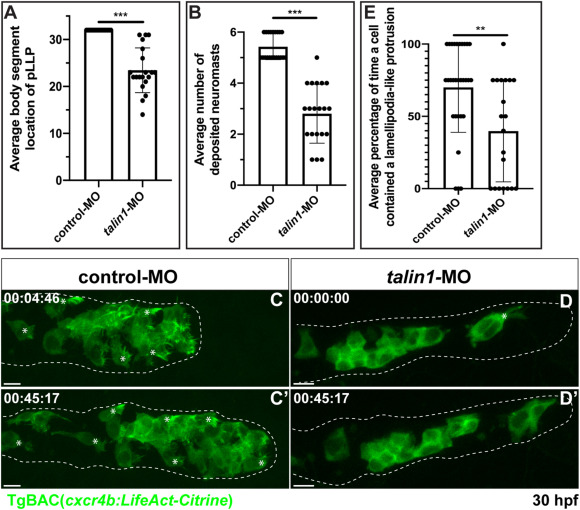
Fig. 4 Fig. 4. Talin1 is necessary for proper lamellipodia-like protrusion formation. (A) Average body segment location of the pLLP 48 hpf in control (0.5 ng) compared to talin1 morpholino (0.5 ng) injected embryos carrying the TgBAC(cxcr4b:LifeAct-Citrine) transgene (n = 20 embryos for each condition). (B) Average number of neuromasts deposited 48 hpf in control-MO and talin-1 MO injected embryos carrying the TgBAC(cxcr4b:LifeAct-Citrine) transgene (n = 20 embryos for each condition). (C and D) Mosaic pLLPs containing cells from TgBAC(cxcr4b:LifeAct-Citrine)-positive embryos that were injected with either 0.5 ng of a control or 0.5 ng of talin1 MOs. Embryos were imaged laterally between 28 and 32 hpf to visualize lamellipodia-like protrusions (Movie 6). (E) Average percentage of time a cell contained a lamellipodia-like protrusion in the control mosaic embryos compared to the talin1 morpholino mosaic embryos. Note that the average percentage of time a cell contained a lamellipodia like protrusion was reduced following Talin1-deficient pLLP cells. Control MO: n = 30 cells from 10 embryos; talin1 MO: n = 20 cells from 8 embryos. Asterisks = lamellipodia-like protrusions. Scale bar = 10 μm.
Reprinted from Developmental Biology, 469, Olson, H.M., Nechiporuk, A.V., Lamellipodia-like protrusions and focal adhesions contribute to collective cell migration in zebrafish, 125-134, Copyright (2020) with permission from Elsevier. Full text @ Dev. Biol.