
|
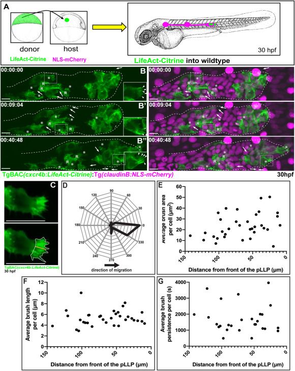
Fig. 1 Fig. 1. Dynamic behavior of brush-like protrusions during pLLP migration. (A) Schematic of the approach used to create mosaic embryos. Cells from TgBAC(cxcr4b:LifeAct-Citrine) embryos were transplanted into embryos positive for Tg(claudinB:NLS-mCherry). (B) Lateral view of stills from a mosaic pLLP imaged at 30 hpf (Movie 1). Insets show brush-like protrusions at 1.5X.(C) Schematic of protrusion parameter measurements: the yellow line and white outline indicate brush length and area, respectively. (D) Orientation of brush-like protrusions in regards to the direction of migration. (E) Average brush area per cell plotted as a function of the location of the cell within the pLLP (0 μm – caudal pLLP tip). (F) Average brush length per cell plotted as a function of the location of the cell within the pLLP. (G) Average brush persistence per cell plotted as a function of the location of the cell within the pLLP. (D–G) Protrusion parameters were measured from 27 to 34 cells in 15–17 chimeric embryos. Brush-like protrusions (asterisk) were defined as those containing ≥2 actin cables (arrowheads). Filopodial protrusions are marked by arrows. Scale bar = 10 μm. R = cells incorporated into rosettes.
Reprinted from Developmental Biology, 469, Olson, H.M., Nechiporuk, A.V., Lamellipodia-like protrusions and focal adhesions contribute to collective cell migration in zebrafish, 125-134, Copyright (2020) with permission from Elsevier. Full text @ Dev. Biol.