
|
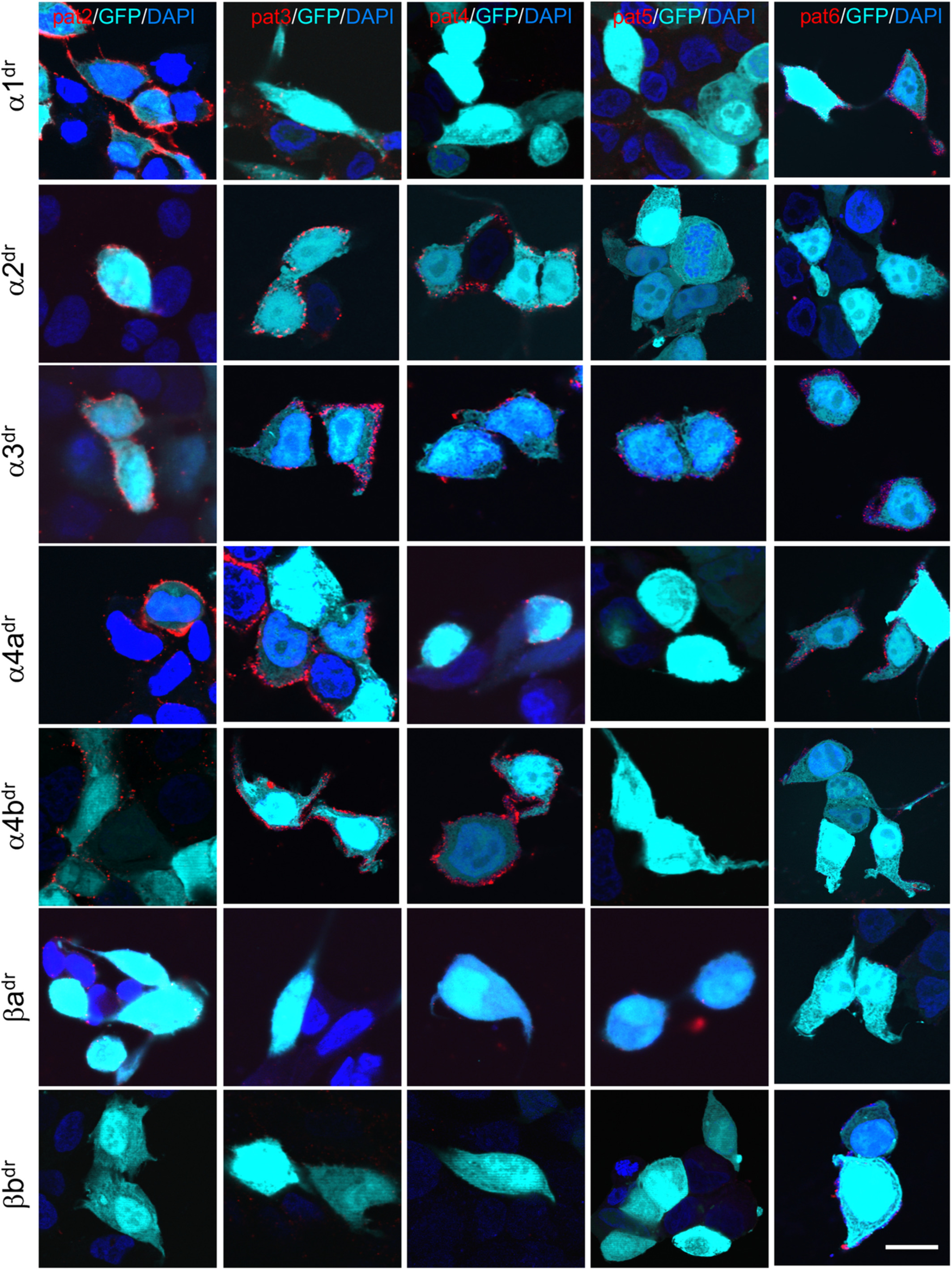
Fig. 7 All patient sera target some GlyR subunits of the zebrafish. HEK293 cells cotransfected with green fluorescent protein (GFP; cyan) and GlyR subunits of Danio rerio (dr; α1, α2, α3, α4a, α4b, βa and βb) were stained with sera from Patient 2 (pat2), pat3, pat4, pat5, and pat6 (red). 4,6‐Diamidino‐2‐phenylindole (DAPI) (blue) labeled the nucleus. White bar in right column represents 20μm. Note that most patient sera target α2, α3, and α4a. The β subunits of D. rerio do not represent a common target for the human GlyR autoantibodies. Stainings for the β subunits were performed following permeabilization of the cells, because the β subunits are not transported to the surface without the presence of α subunits.