
|
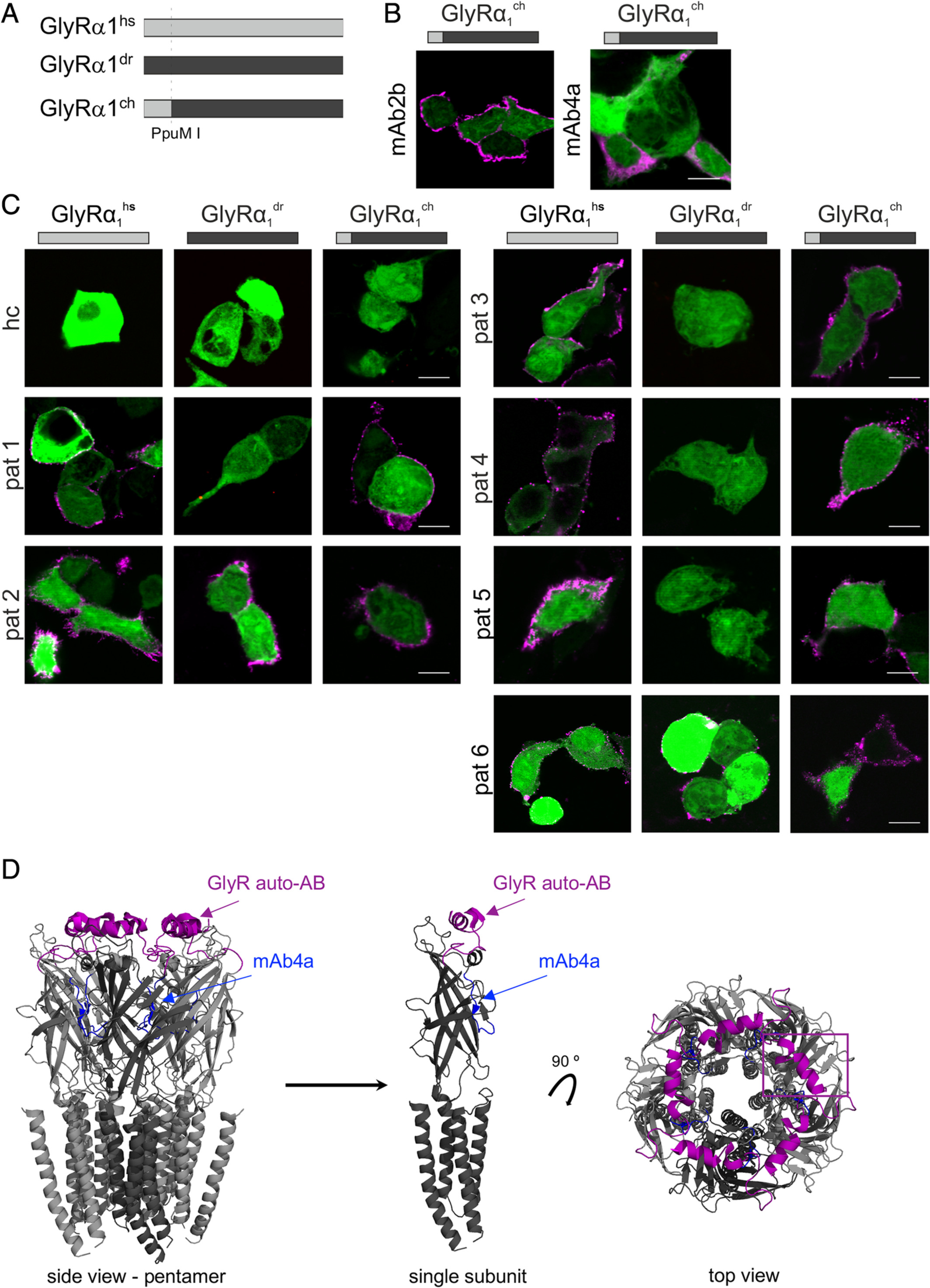
Fig. 5 The GlyRα1 far N‐terminus represents a common epitope for GlyR autoantibodies. (A) Chimeric GlyRα1ch is composed of the N‐terminal part of GlyRα1hs (gray bar) and the C‐terminal part of GlyRα1dr (black bar). Cutting site is indicated by a vertical dotted line (PpuM I, Fig 4A). (B) HEK293 cells where cotransfected with green fluorescent protein (green) and either GlyRα1hs, GlyRα1dr, or GlyRα1ch. GlyRα1ch was detected by the pan‐GlyR antibody mAb4a and mAb2b. (C) Serum of Patient 1 (pat1), pat3, pat4, and pat5 refer to the same staining pattern as mAb2b. The serum from pat2 and pat6 stained all 3 GlyR variants: GlyRα1hs, GlyRα1dr, and GlyRα1ch. Serum from a healthy control served as negative control. White bars = 10μm. (D) Structural model of the GlyRα1 according to Du et al.19 Labeled are the proposed epitope for GlyR autoantibodies (pink) and the mAb4a epitope (blue). Left, pentamer; center, single subunit from side; right, top view on pentamer GlyR. Note the surface localization of the GlyR autoantibody epitope (pink) in contrast to the mAb4a epitope (blue) deeper in the structure at the interface between two adjacent subunits. ch = chimeric; dr = Danio rerio; hs = Homo sapiens.