
|
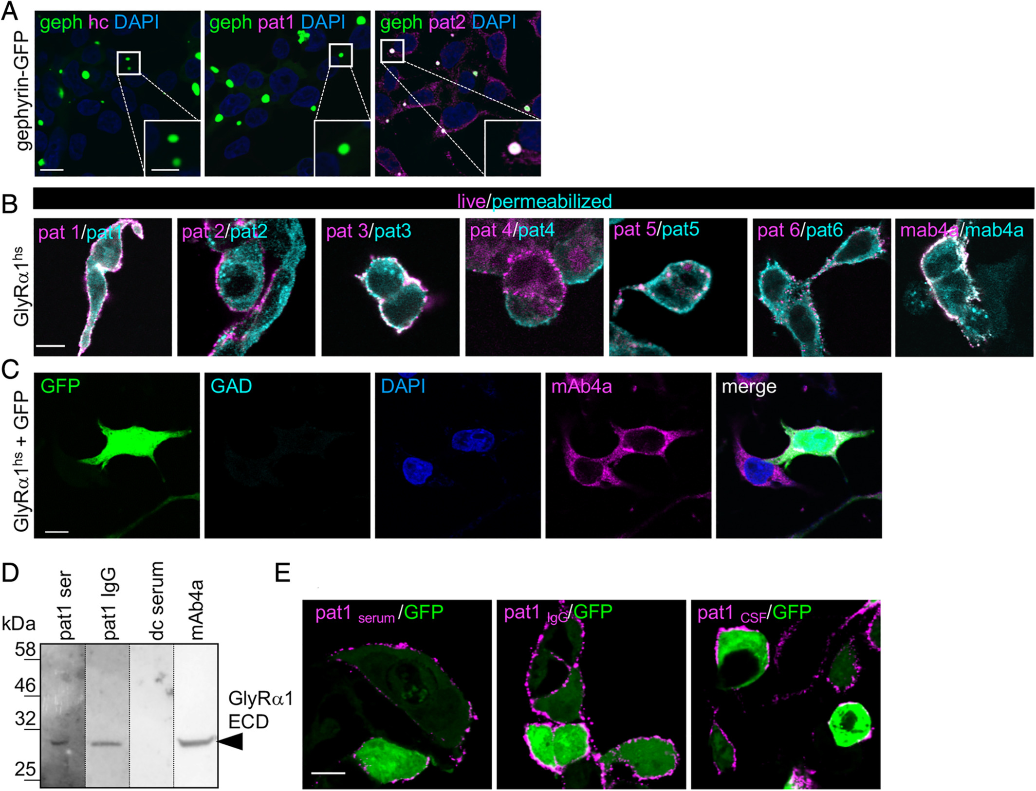
Fig. 1 GlyR‐autoantibodies bind to native and denatured receptor protein. (A) HEK293 cells transfected with a fusion protein of gephyrin and green fluorescent protein (geph‐GFP, green) were stained with healthy control serum (hc), serum of Patient 1 (pat1), and serum of pat2. Intensive colocalization of targeted gephyrin by pat2 autoantibodies and geph‐GFP in the GFP blobs within the cell (see magnified inset, white bar in inset represents 5μm). Positive gephyrin labeling (pink) was also observed in the intracellular part of the cell. Nuclei were marked by 4,6‐diamidino‐2‐phenylindole (DAPI; blue). (B) Transfected HEK293 cells with GlyRα1hs were used. MAb4a (GlyRα1hs fixed and permeabilized) was used to detect GlyRα subunits and served as positive control (right image). Serum GlyR‐autoantibodies from all patients bind to native (magenta) and denatured (cyan) epitopes of GlyRα1hs. (C) Cells were transfected with GFP (green) and GlyRα1hs and stained with glutamic acid decarboxylase (GAD)‐positive control serum (cyan) and the pan‐GlyR antibody mAb4a (pink). Nuclei were marked by DAPI (blue). Scale bars in A–C represent 10μm. (D) The human GlyRα1 extracellular domain (ECD; residues 1–219 = 30.68kDa) was loaded (10μg each lane). The serum of pat1 and the pat1 IgG detected the GlyRα1 ECD similar to the monoclonal antibody mAb4a (pan‐GlyR). dc = disease control, which served as negative control. (E) GlyRα1hs and GFP cotransfected HEK293 cells were incubated with pat1 serum, pat1 purified IgG, and pat1 cerebrospinal fluid (CSF). Magenta fluorescent signals correspond to GlyRα1 bound by the antibodies used. Scale bar = 20μm.