
|
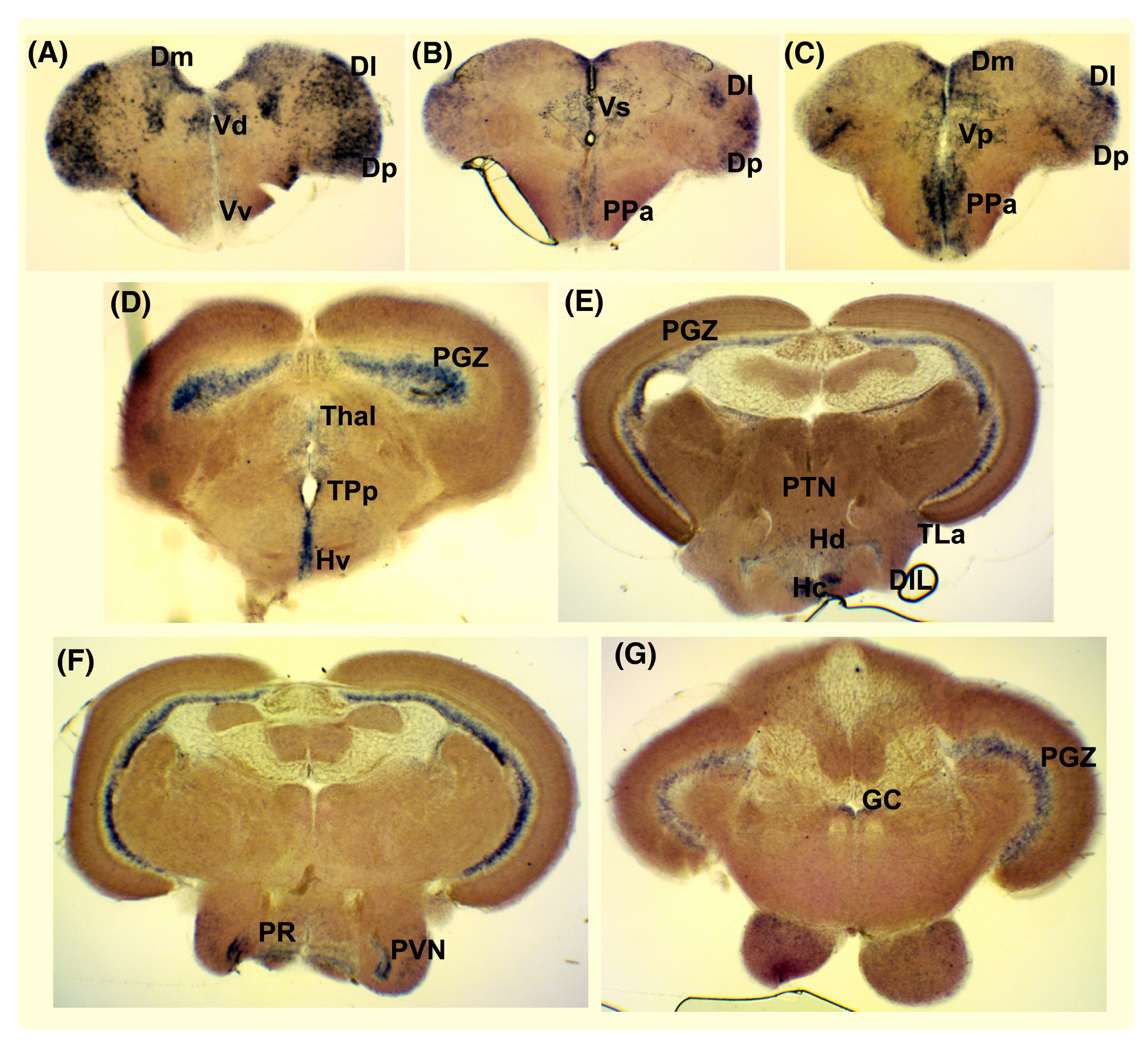
Fig. 1 Expression of histamine receptor H3 (hrh3) in the adult zebrafish brain. A‐G, In situ hybridization showing hrh3 gene expression in the medial, lateral and posterior zone of the dorsal telencephalic area (Dm, Dl and Dp), the ventral, dorsal, supra‐ and post‐commissural nuclei of the ventral telencephalic area (Vv, Vd, Vs and Vp), the anterior parvocellular preoptic nucleus (PPa), the ventral, caudal and dorsal zone of the periventricular hypothalamus (Hv, Hc and Hd), the area around the posterior recess of the diencephalic ventricle (PR), torus lateralis (TLa), periventricular nucleus of posterior tuberculum (TPp), posterior tuberal nucleus (PTN), periventricular nucleus of the inferior lobe of the hypothalamus (PVN), diffuse nucleus of the inferior lobe (DIL), the periventricular grey zone of the optic tectum (PGZ), thalamic nuclei including anterior thalamic nucleus, ventromedial and ventrolateral thalamic nucleus and central and dorsal posterior thalamic nucleus (Thal) and the griseum centrale (GC)