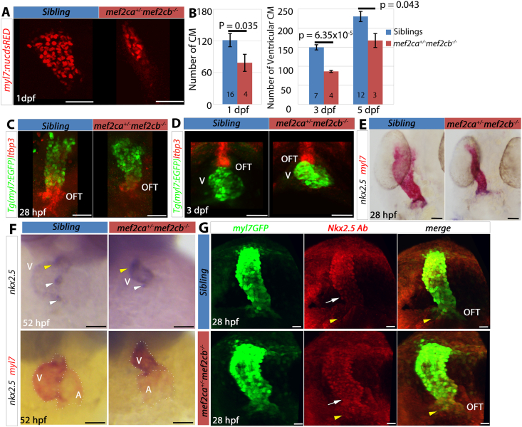
IMAGE

Fig. 5

ID
ZDB-IMAGE-210201-6
Source
Figures for Kula-Alwar et al., 2020
Image
Figure Caption

Fig. 5

Mef2ca+/;mef2cb−/−mutant embryos have reduced CM and changes in SHF gene expression. (A,B) Embryos from a cross of mef2ca+/;mef2cb+/;Tg(myl7:EGFP) and mef2cb+/;Tg(myl7:EGFP);Tg(myl7:nucDsRed) were fixed and immunostained with GFP (green) and RFP (red) antibodies at 1 dpf or imaged live at 3 and 5 dpf and CM nuclei counted. Confocal stacks are shown on red channel only at 1 dpf for clarity (A). Nuclei number of mef2ca+/;mef2cb−/− (red bars) was significantly lower than in siblings (blue bars) at all stages (Unpaired Student’s t-test, P-values indicated on graph). Values are mean ​± ​S.E.M. N shown on bars. (C,D) Embryos from a cross of mef2ca+/;mef2cb+/;Tg(myl7:EGFP) to mef2cb+/ at 28 hpf (C) and 3 dpf (D) hybridised for ltbp3 mRNA (Fast Red) and myl7:EGFP detected with anti-GFP antibody. Hearts of mef2ca+/;mef2cb−/− showed less differentiated myocardium (myl7:EGFP) co-expressing ltbp3 mRNA and ltbp3 levels outside the heart were also reduced (C), but by 3 dpf little difference was seen between genotypes (D). (E,F) In situ mRNA hybridisation for nkx2.5 (blue) and myl7 (red) in embryos from a cross of mef2ca+/;mef2cb+/ and mef2cb−/− at 28 hpf (E, dorsal view) and 52 hpf (F, ventral view, same embryos shown in top panel for nkx2.5 only and in bottom panel for nkx2.5 and myl7). nkx2.5 ​mRNA was upregulated slightly at 28 hpf but was clearly upregulated throughout the ventricle and down in the atrium at 52 hpf, beyond the normal strong expression in AVC (white arrowhead) and the arterial pole (yellow arrowhead). Dotted line (bottom panel, F) marks chambers. (G) Confocal stack of immunodetection of Nkx2.5 and GFP (myl7:EGFP) of hearts of mef2ca+/;mef2cb+/ and sibling at 28 hpf in dorsal view showing Nkx2.5 upregulation in the ventricular region of the heart tube (white arrow) and in the arterial pole (yellow arrowhead). Scale bars ​= ​50 ​μm.

Acknowledgments
This image is the copyrighted work of the attributed author or publisher, and ZFIN has permission only to display this image to its users. Additional permissions should be obtained from the applicable author or publisher of the image.

Reprinted from Developmental Biology, 470, Kula-Alwar, D., Marber, M.S., Hughes, S.M., Hinits, Y., Mef2c factors are required for early but not late addition of cardiomyocytes to the ventricle, 95-107, Copyright (2020) with permission from Elsevier. Full text @ Dev. Biol.