
|
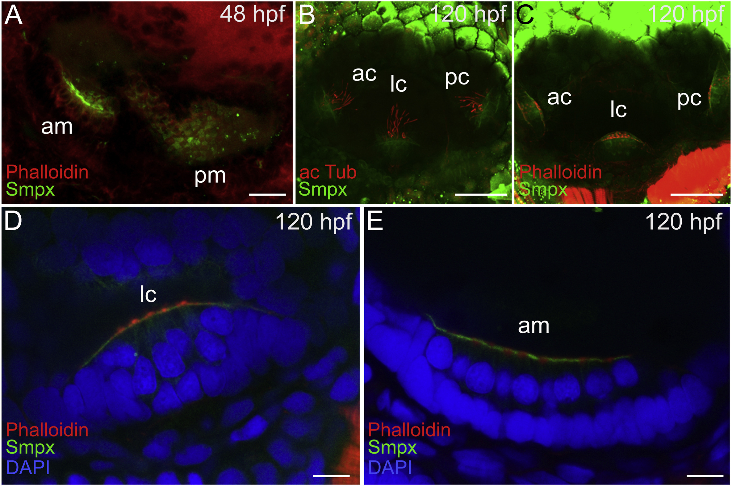
Fig. 3 Smpx embryonic expression pattern analyzed by immunofluorescence. (A) Image of the anti-Smpx antibody staining the ear of a 48 hpf embryo and counterstained with phalloidin (F-actin) to visualize the cytoskeleton. Smpx signal is restricted to the region of the anterior and posterior maculae. (B,C) At 120 hpf Smpx is confined to the anterior, lateral and posterior cristae, labeling the apical membrane of the hair cells, below the kinocilia stained with the antibody against acetylated tubulin (B) and the stereocilia bundle painted with phalloidin (C). (D,E) Close-up views of the zebrafish ear at 120 hpf highlighting the lateral crista and the anterior macula; co-labeling of the apical membrane of the hair cells with the antibody against Smpx and with phalloidin; the nuclei (DAPI) are located in the basal portion of the cells. Images are all lateral views, anterior to the left, of confocal Z-stacks taken from whole mount embryos and larvae. am, anterior macula; pm, posterior macula; ac, anterior crista; lc, lateral crista; pc, posterior crista. Scale bars = 20 μm in A; 50 μm in B,C; 10 μm in D,E.
Reprinted from Gene expression patterns : GEP, 36, Ghilardi, A., Diana, A., Prosperi, L., Del Giacco, L., Expression pattern of the small muscle protein, X-linked (smpx) gene during zebrafish embryonic and larval developmental stages, 119110, Copyright (2020) with permission from Elsevier. Full text @ Gene Expr. Patterns