Image
Figure Caption

Figure 1—figure supplement 2. Electron microscopy analysis of endosomes in the absence of RalA or RalB (<bold>a-b</bold>).

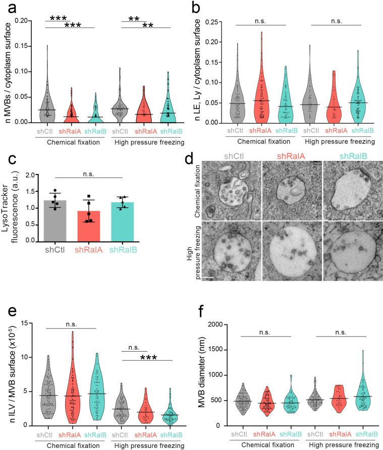
Graph showing the number of multi-vesicular body (MVB) (a) or endolysosomes (b) per cytoplasm surface in electron microscopy analysis performed by chemical fixation or high-pressure freezing on 4T1 shControl, shRalA, and shRalB cells. Each dot represents one field of view. Kruskal-Wallis test followed by Dunn's Multiple Comparison Test. (c) FACS analysis of 4T1 shControl, shRalA, and shRalB cells stained with Lysotracker. Each dot represents one experiment (five independent experiments; Kruskal-Wallis test followed by Dunn's Multiple Comparison Test). (d) Representative electron micrographs of MVBs in 4T1 shControl, shRalA, and shRalB cells after chemical fixation or high-pressure freezing. Scale bar: 200 nm. (e–f) Number of ILV per MVB surface (e) and MVB diameter (f) measured in electron microscopy analysis performed by chemical fixation or high-pressure freezing on 4T1 shControl, shRalA, and shRalB cells. Each dot represents one MVB; Kruskal-Wallis test followed by Dunn's Multiple Comparison Test.

Acknowledgments
This image is the copyrighted work of the attributed author or publisher, and ZFIN has permission only to display this image to its users. Additional permissions should be obtained from the applicable author or publisher of the image. Full text @ Elife