IMAGE

FIGURE 1

ID
ZDB-IMAGE-210128-20
Source
Figures for Zhu et al., 2021
Image
Figure Caption

FIGURE 1

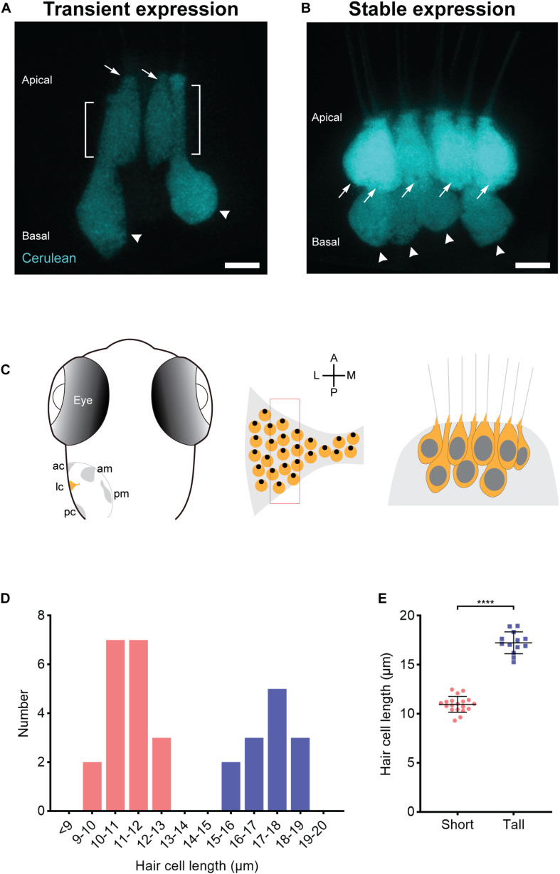
Two populations of morphologically distinct hair cells in the central thickness of the lateral crista of larval zebrafish. (A,B) Transient (A) and stable (B) transgenic expression of Cerulean fluorescent protein in the lateral crista reveals two morphologically distinct hair cells: short (arrows) and tall (arrowheads). The brackets mark the neck regions of tall hair cells. Scale bar = 5 μm. (C) Left panel, schematic of a dorsal view of a 7-dpf zebrafish ear, illustrating the five mechanosensory patches: the anterior crista (ac), lateral crista (lc), posterior crista (pc), anterior macula (am), and posterior macula (pm). Middle panel, enlarged view schematizing the hair cell arrangement in the lateral crista highlighted in yellow in the left panel. Each yellow circle represents a single hair cell, and the black dot within represents the position of the kinocilium (fonticulus). The red rectangle marks the section from which the hair cells are imaged and characterized. A, anterior; P, posterior; M, medial; L, lateral. Right panel, schematic of the two populations of hair cells (short and tall) in this section of the lateral crista based on imaging results. (D) The distribution of somata lengths of the lateral crista hair cells from 7-dpf zebrafish. n = 4. Pink bars represent short hair cells. Blue bars represent tall hair cells. (E) The somata lengths (mean ± SD) of the two groups of hair cells shown in panel (D). ****P < 0.0001, unpaired t-test.

Acknowledgments
This image is the copyrighted work of the attributed author or publisher, and ZFIN has permission only to display this image to its users. Additional permissions should be obtained from the applicable author or publisher of the image. Full text @ Front Cell Dev Biol