
|
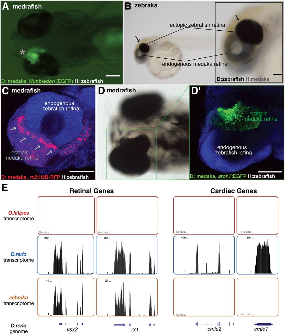
Fig. 3 Transplanted EGFP+ cluster develops into an ectopic retina both in zebraka and medrafish. Images of medrafish (A,C,D) and zebraka (B). The EGFP+ cluster (asterisk, ventral view of a hatch embryo; A) and the pigmented cluster (arrows, lateral view of a hatched embryo; B) develop into an ectopic retina (n=45 transplantation events). Confocal images show expression markers of retinal progenitors (arrows in C; rx2:H2B-RFP donors in medrafish at 4 dpf) and retinal neurogenesis (D; atoh7:EGFP donors in medrafish at 5 dpf) (C,D′, DAPI in blue) (n=17 transplantation events). (E) Transcriptomes of medaka (top, n=2), zebrafish (middle, n=2) and zebraka (bottom, n=3) plotted along the zebrafish genome. The zebrafish cells in medrafish display retinal identity (vsx2 and rx1, left two panels) and no cardiac gene markers (cmlc1 and cmlc2, right panels). Scale bars: 100 µm. D, donor; H, host.