
Image
|
Figure Caption
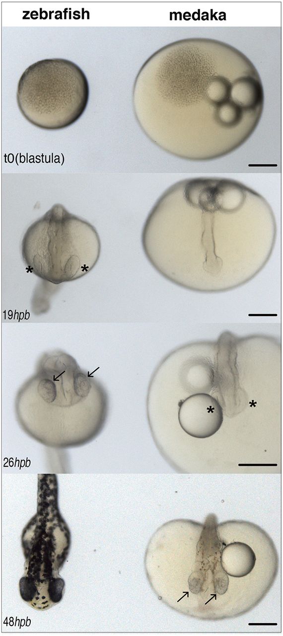
Fig. 1 Different developmental timing for zebrafish and medaka. Images of zebrafish (left) and medaka (right) embryos synchronised at the blastula stage (top, 512 cells, t=0) and at different stages of embryonic development (hours post blastula, hpb). Eye cups (asterisks) are evident in zebrafish by 19 hpb and retinal pigmentation (and neurogenesis) (arrows) by 26 hpb; in medaka, retinal pigmentation does not start before 48 hpb. Scale bars: 200 µm.
Acknowledgments
This image is the copyrighted work of the attributed author or publisher, and
ZFIN has permission only to display this image to its users.
Additional permissions should be obtained from the applicable author or publisher of the image.
Full text @ Development