IMAGE

Figure 4

ID
ZDB-IMAGE-210113-148
Antibodies
Source
Figures for Sun et al., 2020
Image
Figure Caption

Figure 4

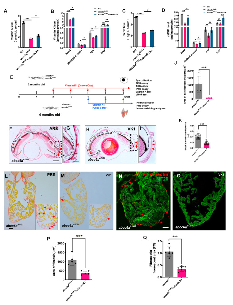
Vitamin K treatment relives ocular calcification and cardiac fibrosis in abcc6aΔ1/Δ1 mutants. (AD) Graphical representation of sandwich ELISA measurements for tissue and serum vitamin K and cMGP levels in WT (n = 18) and DMSO/ethanol- (n = 18) or vitamin K1-treated abcc6aΔ1/Δ1 fish (n = 18) fish. * p < 0.05, ** p < 0.01, *** p < 0.001, **** p < 0.0001, Student’s t-test (unpaired, two-tailed). (E) Experimental design for once-a-day DMSO/ethanol (0.1%) and vitamin K1 (80 µM) treatment of abcc6a+/+ and abcc6aΔ1/Δ1 fish. Red arrows indicate experimental steps, changing water every day for 4 months from 2 months old; for eye collection, TEM, ARS, and PRS assay, vitamin K and cMGP tests. Blue arrows indicate experimental steps for heart collection, PRS assay and immunostaining analyses for 2 months from 4 months of age. (FI) Vitamin K rescues abnormal scleral calcification in abcc6aΔ1/Δ1 mutant zebrafish. (G,I) Higher-magnification images of the dashed boxes in (F,H). Dashed black line indicates approximate plane of resection. Red arrow marks abnormal calcification of scleral layer in the abcc6aΔ1/Δ1 mutant eyes. (J) Quantification of area of calcification of sclera after 4 months of DMSO/ethanol (n = 7) or vitamin K1 treated treatment in abcc6aΔ1/Δ1 fish (n = 7). *** p < 0.001, Student’s t-test (unpaired, two-tailed). (K) Quantification of Bruch’s membrane thickness after 4 months of DMSO/ethanol (n = 13, 8 measuring points per sample) or vitamin K1 treatment in abcc6aΔ1/Δ1 fish (n = 15, 8 measuring points per sample). *** p < 0.001, Student’s t-test (unpaired, two-tailed). (L,M) Picrosirius Red staining of heart sections shows significantly reduced collagen deposition reside in the compact layer and trabeculae after treatment with vitamin K1 (M), as compared to abcc6aΔ1/Δ1 mutant hearts after treatment with DMSO/ethanol (L). Red for collagen (L; Red arrow) and yellow for cardiomyocyte fiber. (N,O) Immunofluorescent section images of adult ventricles stained with anti-Fibronectin and anti-MF20 antibodies from DMSO/ethanol-treated (N) or vitamin K1 treated abcc6aΔ1/Δ1 mutant fish (O), showing that vitamin K reduces Fibronectin. (P) Quantification of area of fibrosis after 2 months of DMSO/ethanol (n = 5) or vitamin K1 treatment in abcc6aΔ1/Δ1 fish (n = 5). *** p < 0.001, Student’s t-test (unpaired, two-tailed). (Q) Quantification of Fibronectin fluorescent area after 2 months of DMSO/ethanol (n = 7) or vitamin K1 treatment in abcc6aΔ1/Δ1 fish (n = 7). *** p < 0.001, Student’s t-test (unpaired, two-tailed). Scale bar: 250 μm (F,H); 50 μm (LO); 125 μm (G,I).

Figure Data
Acknowledgments
This image is the copyrighted work of the attributed author or publisher, and ZFIN has permission only to display this image to its users. Additional permissions should be obtained from the applicable author or publisher of the image. Full text @ Int. J. Mol. Sci.