IMAGE

Fig 6

ID
ZDB-IMAGE-210113-123
Source
Figures for Nunley et al., 2020
Image
Figure Caption

Fig 6 Cone mosaic formation is minimally disrupted in Red cone mutant but is significantly disrupted in UV cone mutant.

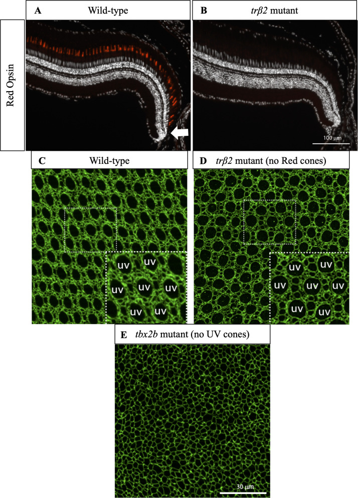
(A) Cross-section of wild-type retina in which immunostaining of Red cone opsin labels Red cones. White arrow indicates approximate location of precolumn area [40]. (B) Cross-section of trβ2 mutant in which immunostaining of Red cone opsin labels Red cones. Note absence of differentiated Red cones. White arrow indicates approximate location of precolumn area. (C) Apical plane of wild-type cone mosaic lattice in retinal flat-mount in which anti-ZO-1 stains cell profiles. UV cones are indicated in the inset. (D) Apical plane of cone mosaic in retinal flat-mount from trβ2 mutant, which lacks Red cones. Anti-ZO-1 stains cell profiles. UV cones (indicated in inset) identified based on large, rounded profiles. The triangular lattice of UV cones is minimally disrupted in absence of Red cones. (E) Apical plane of cones in retinal flat-mount from tbx2b mutant, which lacks UV cones. The cone mosaic is disrupted in this mutant.

Figure Data
Acknowledgments
This image is the copyrighted work of the attributed author or publisher, and ZFIN has permission only to display this image to its users. Additional permissions should be obtained from the applicable author or publisher of the image. Full text @ PLoS Comput. Biol.