IMAGE

Figure 3

ID
ZDB-IMAGE-210113-12
Source
Figures for Danesin et al., 2021
Image
Figure Caption

Figure 3

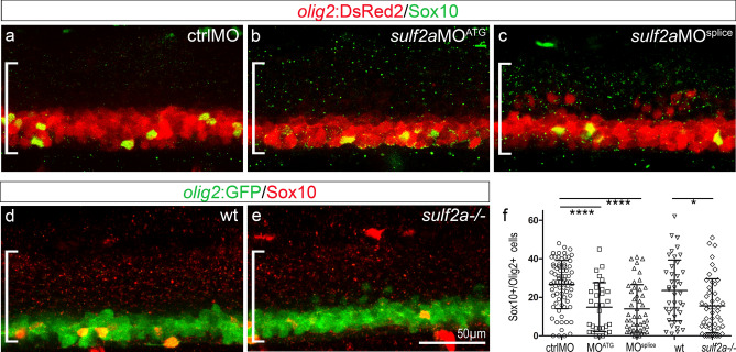
Sulf2a depletion impairs OPC development. Side views of 48 hpf embryos. (a-f) Detection (a–e) and quantification (f) of OPCs by immunodetection of Sox10 (green in a–c, red in d,e) in Tg(olig2:DsRed) embryos (red in a–c) injected with ctrlMO (a, n = 73), sulf2aMOATG (b, n = 31) or sulf2aMOsplice (c, n = 47) and wild-type (d, n = 10) or sulf2a−/− (e, n = 48) Tg(olig2:GFP) embryos (green in d,e) from 9 and 6 independent experiments, respectively. Datasets were compared with Mann Whitney’s test (two-tailed). Data are presented as mean number of cells per embryo ± s.d (*p < 0.05, ****p < 0.0001).

Figure Data
Acknowledgments
This image is the copyrighted work of the attributed author or publisher, and ZFIN has permission only to display this image to its users. Additional permissions should be obtained from the applicable author or publisher of the image. Full text @ Sci. Rep.