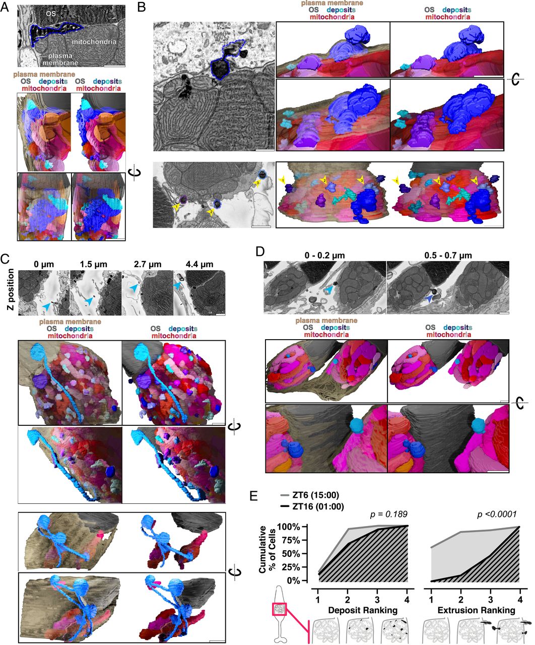
Fig. 5 Mitochondria share material and extrude it from the cell in darkness. (A) SBFSEM image (Top) of deposits associated with cone mitochondria and corresponding 3D renderings (Bottom). One large deposit (blue outline) below the OS is associated with multiple mitochondria. (B) SBFSEM images showing extrusion of mitochondrial-associated deposits from cones, with 3D renderings. (B, Top) Extrusion of one deposit (blue outline). (B, Bottom) Multiple extrusion events in one cell (blue-violet outlines, yellow arrows). (C) SBFSEM images showing stalks and networks in the extracellular space, and corresponding 3D renderings. (C, Middle) One extruded deposit tethered to mitochondria by a stalk (blue arrows). (C, Bottom) Branched network of extruded material contacting three distinct populations of cone mitochondria from one cell. (D) Extrusion of mitochondrial-associated deposits from two neighboring rods. (D, Top) SBFSEM minimum intensity projections over 0.2-µm depth highlighting extrusion events (blue arrows). (D, Bottom) Three-dimensional rendering. (All scale bars, 1 µm.) Beige, plasma membrane; gray, OSs; reds, mitochondria; blues, deposits. (E) Quantification of deposits and extrusion events at ZT6 (gray) and ZT16 (black). For deposits: 1 (no mitochondrial deposits) to 4 (every mitochondrion having a deposit). For the number of extrusion events: 1 (no events) to 4 (more than three events). SI Appendix, Table S2 lists Ns from all groups.
Image
Figure Caption
Acknowledgments
This image is the copyrighted work of the attributed author or publisher, and
ZFIN has permission only to display this image to its users.
Additional permissions should be obtained from the applicable author or publisher of the image.
Full text @ Proc. Natl. Acad. Sci. USA

