IMAGE

Fig. 5.

ID
ZDB-IMAGE-201231-9
Source
Figures for Koleilat et al., 2020
Image
Figure Caption

Fig. 5.

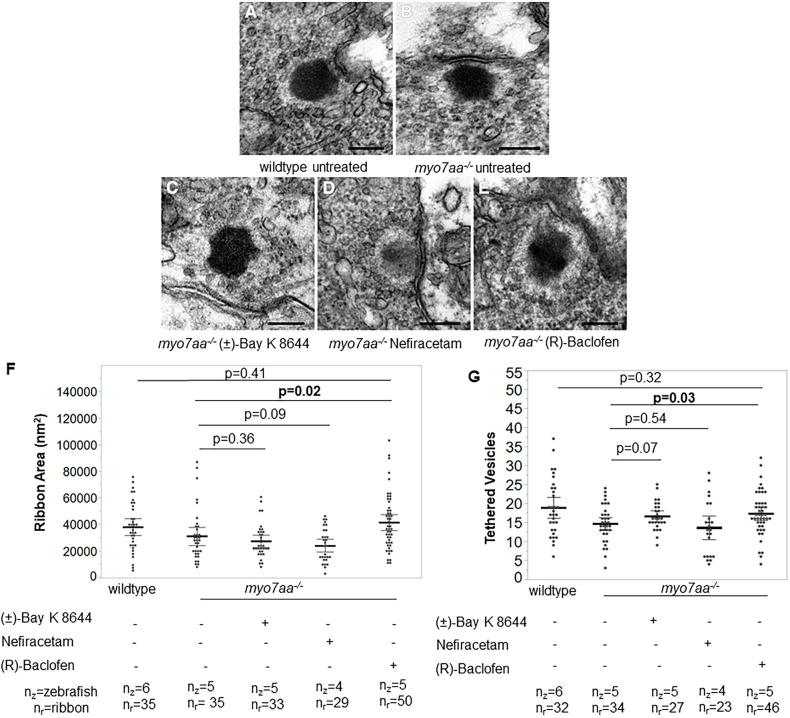
myo7aa−/− mutant ribbon synapse abnormalitiesarediminishedafterexposure to (R)-Baclofen. (A-E) Representative TEM images showing ribbon synapses of untreated wild-type (A) and untreated myo7aa−/− mutant (B) larvae, as well as myo7aa−/− mutants treated with 5 µM (±)-Bay K 8644 (C), 250 µM Nefiracetam (D) or 125 µM (R)-Baclofen (E). All images were taken at 5 dpf. Scale bars: 1000 nm. (F) The ribbon area in untreated myo7aa−/− is smaller compared to that in myo7aa−/− treated with 125 µM (R)-Baclofen. Ribbon synapses of untreated wild-type larvae have a ribbon area comparable to that of myo7aa−/− mutants treated with 125 µM (R)-Baclofen (two-tailed t-test). (G) Ribbons of untreated myo7aa−/− mutants have fewer tethered vesicles compared to those of myo7aa−/− mutants treated with 125 µM (R)-Baclofen. Ribbons of untreated wild-type larvae have a number of tethered vesicles comparable to those of myo7aa−/− larvae treated with 125 µM (R)-Baclofen (two-tailed t-test). Bold black lines represent the mean of the data set and error bars are 95% confidence intervals. Experiments were replicated three times for myo7aa−/− mutants treated with each drug.

Figure Data
Acknowledgments
This image is the copyrighted work of the attributed author or publisher, and ZFIN has permission only to display this image to its users. Additional permissions should be obtained from the applicable author or publisher of the image. Full text @ Dis. Model. Mech.