IMAGE

FIGURE 2

ID
ZDB-IMAGE-201230-2
Source
Figures for Delbaere et al., 2020
Image
Figure Caption

FIGURE 2

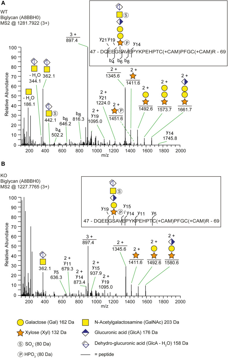
The identification of a trisaccharide linkage region in the b3galt6–/– zebrafish. (A,B) MS2 fragment mass spectra of CS-glycopeptides derived from biglycan (UniProt: A8BBH0) showed different linkage regions in WT compared to the b3galt6–/– zebrafish. (A) The peptide (DQEEGSAVEPYKPEHPTCPFGCR) in WT zebrafish was modified with a residual hexasaccharide structure, corresponding to the canonical tetrasaccharide linkage region (m/z 1281.79; 3+). The hexasaccharide structure was modified with one sulfate group (SO 3-) at the subterminal GalNAc residue and one phosphate group (HPO3-) at the xylose residue (Supplementary Figure S7). (B) The peptide (DQEEGSAVEPYKPEHPTCPFGCR) in b3galt6–/– zebrafish was modified with a residual pentasaccharide structure, corresponding to a non-canonical trisaccharide linkage region (m/z 1227.78; 3+). The pentasaccharide structure was modified with two phosphate/sulfate groups. Their exact positioning and identity could not be determined due to low intensities of corresponding ion peaks, and the positions are therefore assigned in analogy of the WT structure.

Acknowledgments
This image is the copyrighted work of the attributed author or publisher, and ZFIN has permission only to display this image to its users. Additional permissions should be obtained from the applicable author or publisher of the image. Full text @ Front Cell Dev Biol