IMAGE

Figure 1

ID
ZDB-IMAGE-201229-107
Source
Figures for Liedtke et al., 2020
Image
Figure Caption

Figure 1

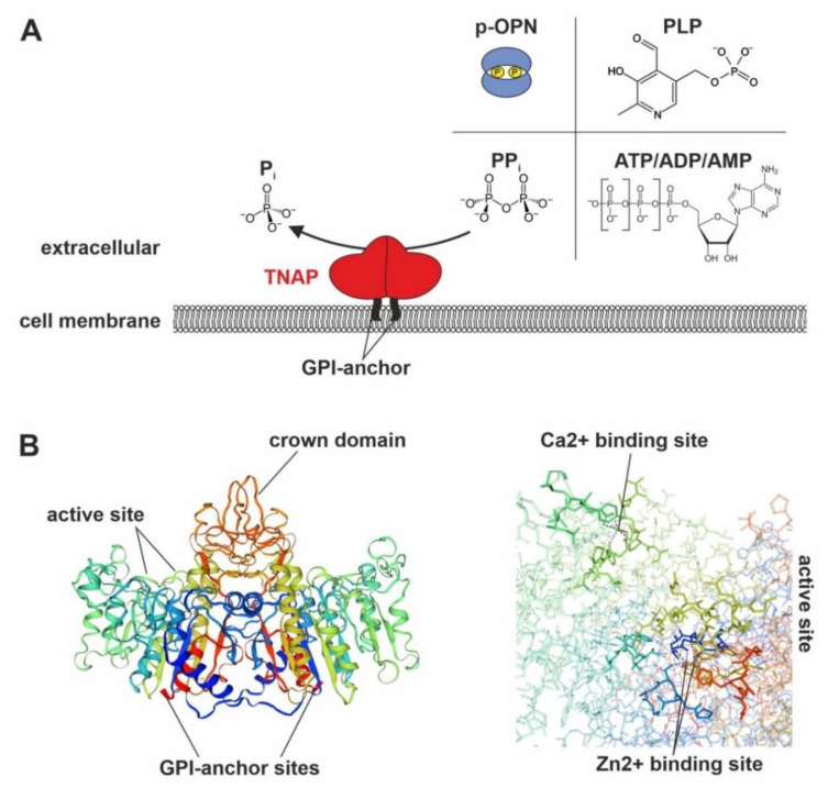
Potential enzymatic functions and structural visualization of TNAP. (A) Depiction of potential TNAP substrates. (B) Schematic depiction of a predicted homology model of TNAP’s dimerized 3D structure (https://swissmodel.expasy.org/; SWISS-MODEL: P05186 (PPBT_HUMAN); homology model according to the template: 3mk1.1.B “Refinement of placental alkaline phosphatase complexed with nitrophenyl”) and magnified view of the active site [26,27,28]. Figure 1B was modified according to SWISS-MODEL database terms of use and is licensed under a Creative Commons Attribution 4.0 (https://creativecommons.org/licenses/by-sa/4.0/). ATP/ADP/AMP: adenosine tri-/di-/monophosphate, GPI-anchor: glycosyl-phosphatidylinositol anchor, Pi: phosphate, PLP: pyridoxal-5′-phosphate, p-OPN: phosphorylated osteopontin, PPi: inorganic pyrophosphate, TNAP: tissue-nonspecific alkaline phosphatase.

Acknowledgments
This image is the copyrighted work of the attributed author or publisher, and ZFIN has permission only to display this image to its users. Additional permissions should be obtained from the applicable author or publisher of the image. Full text @ Biomolecules