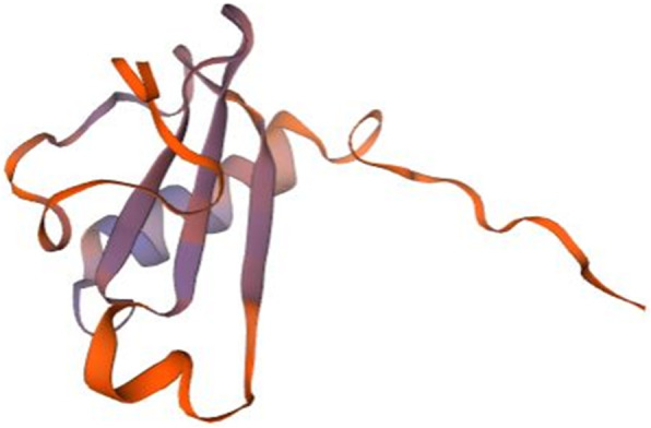
IMAGE

Fig. 4

ID
ZDB-IMAGE-201212-16
Source
Figures for Issaka Salia et al., 2020
Image
Figure Caption

Fig. 4

Homology model of P10 chemokine interleukin-8-like domain. Lymphotactin (1j8i.1.A in the rcsb protein database) is the template used for the homology modelling of P10. The homology model starts from P10 residue N°24 (GLU, Glutamic acid) and ends with the residue N° 102 (SER, Serine). The NMR spectroscopy was used to determine the experimental structure of 1j8i.1.A [62]. The blue color show regions of the model where P10 was well modeled and orange regions where P10 was poorly modeled. The chemokine interleukin-8-like domain of the model starts with P10 amino acid at position N°27(HIS, Histidine) and ends with amino acid at position N°86 ((LEU, Leucine). This region includes both well-modeled (blue) and poorly-modeled (orange) sections

Acknowledgments
This image is the copyrighted work of the attributed author or publisher, and ZFIN has permission only to display this image to its users. Additional permissions should be obtained from the applicable author or publisher of the image. Full text @ BMC Genomics