
IMAGE
Figure 4—figure supplement 1.
- ID
- ZDB-IMAGE-201202-27
- Source
- Figures for Dalle Nogare et al., 2020
Image
|
Figure Caption
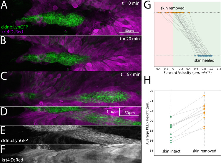
Figure 4—figure supplement 1.
Top panel shows control (unmanipulated) and bottom panel shows experimental (skin removed) side of the same embryo. Images are maximum-intensity projected stitched confocal stacks overlaid with DIC images.
Acknowledgments
This image is the copyrighted work of the attributed author or publisher, and
ZFIN has permission only to display this image to its users.
Additional permissions should be obtained from the applicable author or publisher of the image.
Full text @ Elife