IMAGE

Figure 2

ID
ZDB-IMAGE-201130-51
Genes
Source
Figures for Kakouri et al., 2020
Image
Figure Caption

Figure 2

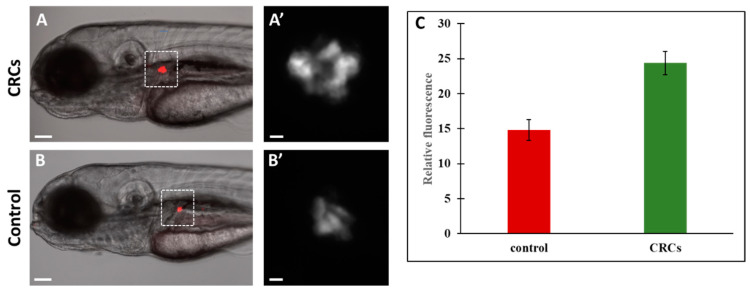
Zebrafish pancreas monitoring after treatment with CRCs. Transgenic zebrafish Tg(ins:DsRed) were treated at 72hpf with 0.2 mg/mL CRCs for 48h and visualized under fluorescent microscope. There was significant increase of fluorescence on the pancreatic islets of CRCs-treated embryos (A,A′) compared to the control group (B,B′). A’ and B’ are micrographs of A and B, respectively. (C) fluorescence intensity was quantified using Fiji software. Results shown are the mean +/− SEM. p < 0.01. Scale bars: A and B: 100 μm, A’ and B’:10 μm.

Figure Data
Acknowledgments
This image is the copyrighted work of the attributed author or publisher, and ZFIN has permission only to display this image to its users. Additional permissions should be obtained from the applicable author or publisher of the image. Full text @ Molecules