IMAGE

Fig 7

ID
ZDB-IMAGE-201119-7
Source
Figures for Rajan et al., 2020
Image
Figure Caption

Fig 7 Model of perivascular fibroblasts in vascular stabilization in zebrafish.

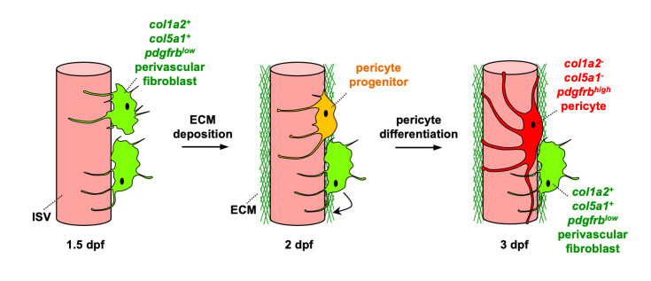
Perivascular fibroblasts, characterized by the expression of fibrillar collagens col1a2 and col5a1 as well as low levels of pdgfrb, become associated with intersegmental vessels (ISVs) in the zebrafish trunk by 1.5 dpf. Perivascular fibroblasts deposit a network of collagen fibers to establish the vascular ECM and stabilize nascent ISVs at 2 dpf and continue to deposit collagen until at least 3 dpf. In the same time window, a subset of perivascular fibroblasts functions as pericyte progenitors. They gradually upregulate the expression of the classic pericyte marker pdgfrb and develop elongated cellular processes. By 3 dpf, these ‘pericyte progenitors’ have completely differentiated into mature pericytes, showing robust pdgfrb expression distinct from collagen-expressing perivascular fibroblasts. Together, perivascular fibroblasts perform dual functions in vascular stabilization by depositing collagens to support nascent blood vessels and acting as pericyte progenitors.

Acknowledgments
This image is the copyrighted work of the attributed author or publisher, and ZFIN has permission only to display this image to its users. Additional permissions should be obtained from the applicable author or publisher of the image. Full text @ PLoS Genet.