IMAGE

Fig 1

ID
ZDB-IMAGE-201119-3
Genes
Source
Figures for Rajan et al., 2020
Image
Figure Caption

Fig 1 Characterization of perivascular fibroblasts in zebrafish.

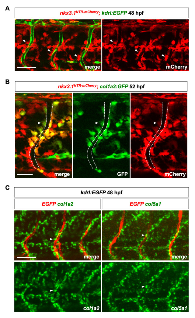
(A) Lateral view of a three somite region in nkx3.1NTR-mCherry; kdrl:EGFP embryos at 48 hpf. Many mCherry+ perivascular fibroblasts (red, arrowheads) were closely associated with intersegmental vessels (ISVs) labeled by the endothelial marker kdrl:EGFP (green). n = 11 embryos. (B) Co-expression of nkx3.1NTR-mCherry and col1a2:GFP in perivascular fibroblasts (arrowheads) at 52 hpf. The ISV is indicated by dashed lines. Graph showing proportion of perivascular fibroblasts labeled by col1a2:GFP is shown in S1A Fig. n = 17 embryos. (C) Fluorescent mRNA in situ hybridization showing expression of fibrillar collagens col1a2 (green, left) and col5a1 (green, right) in perivascular fibroblasts (arrowheads) along ISVs marked by kdrl:EGFP (red) at 48 hpf. n = 15 embryos per staining. Scale bars: (A,C) 50 μm; (B) 25 μm.

Figure Data
Acknowledgments
This image is the copyrighted work of the attributed author or publisher, and ZFIN has permission only to display this image to its users. Additional permissions should be obtained from the applicable author or publisher of the image. Full text @ PLoS Genet.