IMAGE

Figure 5.

ID
ZDB-IMAGE-201119-12
Source
Figures for Beckwith-Cohen et al., 2020
Image
Figure Caption

Figure 5.

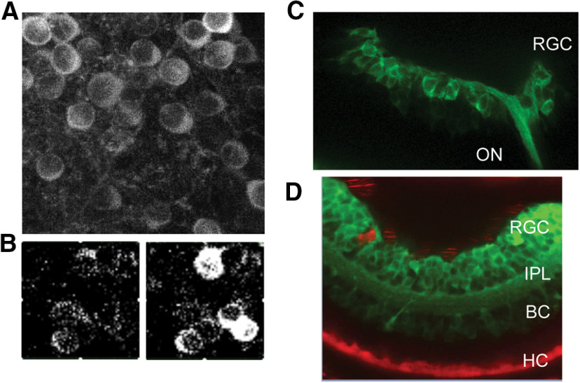
Expression of GCaMP6f in chemogenetic zebrafish lines. A, Ex vivo retina of adult HuC-GCaMP6f zebrafish shows eGFP fluorescence in retinal flat mounts using two-photon imaging. B, Light response is easily measured in RGCs before (left) and after (right) a light flash. C, In vivo confocal imaging of eGFP in 1 dpf PTU-treated zebrafish larvae of HuC-GCaMP6f and FaNaC crossed transgenic fish. The optic nerve (ON) and the developing retina (RGC) show strong eGFP fluorescence. D, In vivo imaging of the same line of fish imaged in C showing that FaNaC-mCherry is easily visualized in HCs at 2 dpf zebrafish larvae. Some red autofluorescence is generated by scattered pigment cells on the ocular surface. RGC, retinal ganglion cell layer; IPL, inner plexiform layer; BC, bipolar cell layer; HC, horizontal cell layer.

Acknowledgments
This image is the copyrighted work of the attributed author or publisher, and ZFIN has permission only to display this image to its users. Additional permissions should be obtained from the applicable author or publisher of the image. Full text @ eNeuro