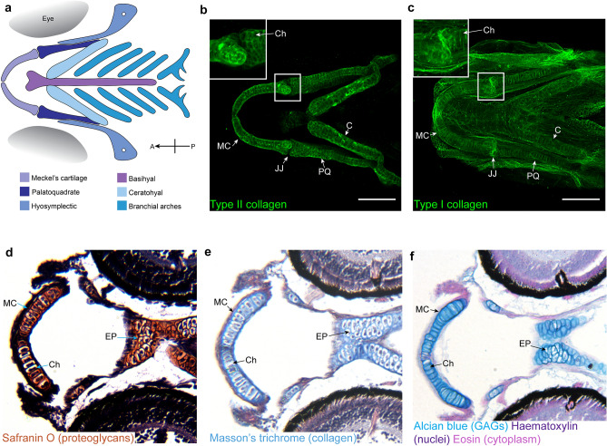
Zebrafish craniofacial cartilage has key components found in human articular cartilage. a Schematic representation of the cartilage elements which make up the zebrafish craniofacial skeleton at 5 dpf. Orientation compass included in bottom right, with A anterior and P posterior. b, c Ventral view confocal image stacks of 5 dpf craniofacial skeleton immunostained for type II collagen (b) and type I collagen (c) in the ECM surrounding chondrocytes. Inset shows zoom of jaw joint. Scale bar = 100 μm. Images in b and c previously published in (Lawrence et al. 2018). d–f Ventral paraffin sections of 5 dpf craniofacial skeleton stained with Safranin O (d) to show proteoglycans, Masson’s trichrome (e) to show the presence of collagen, and alcian blue (f) to show the presence of glycosaminoglycans in the chondrocyte ECM which makes up the cartilage. MC Meckel’s cartilage, JJ jaw joint, PQ palatoquadrate, C ceratohyal, Ch example of a chondrocyte surrounded by ECM, EP ethmoid plate
Acknowledgments
This image is the copyrighted work of the attributed author or publisher, and
ZFIN has permission only to display this image to its users.
Additional permissions should be obtained from the applicable author or publisher of the image.
Full text @ Histochem. Cell Biol.
Your Input Welcome
Thank you for submitting comments. Your input has been emailed to ZFIN curators who may contact you if
additional information is required.
Oops. Something went wrong. Please try again later.