IMAGE

Fig. 2.

ID
ZDB-IMAGE-201102-6
Antibodies
Source
Figures for Jewett et al., 2020
Image
Figure Caption

Fig. 2.

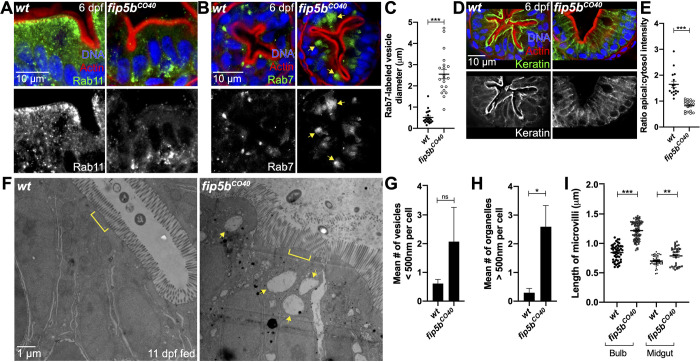
Endosome maturation and terminal web keratin organization requirefip5bfunction. All following images are representative cross sections through the midgut region. Wild-type siblings are used as controls. For all experiments, three separate animals for each condition were analyzed. (A,B,D) Immunohistochemistry on cross sections of 6 dpf wild-type and Fip5bCO40 mutant larvae stained with Hoechst (blue), Phalloidin (red), and Rab11 (A), Rab7 (B), or Cytokeratin (D) (green). (C) Quantitation of Rab7-vesicle diameter. (E) Ratio of fluorescence intensity of apical keratin to cytoplasmic keratin. (F) Electron micrographs showing 11 dpf fed wild-type and fip5bCO40 mutant larvae. Arrows point to larger than 500 nm organelles and brackets mark terminal web. (G) Quantitation of less than 500 nm apical vesicles in 11 dpf larvae. (H) Quantitation of greater than 500 nm organelles in 11 dpf larvae. (I) Quantitation of microvilli length in the intestinal bulb and midgut of 11 dpf larvae. Each dot represents a single microvillus length combined across three animals. All plots show mean±s.e.m. A t-test was used for Gaussian data and a Mann–Whitney test for all other statistics. ***P<0.0005, **P<0.005, *P<0.05.

Figure Data
Acknowledgments
This image is the copyrighted work of the attributed author or publisher, and ZFIN has permission only to display this image to its users. Additional permissions should be obtained from the applicable author or publisher of the image. Full text @ Biol. Open