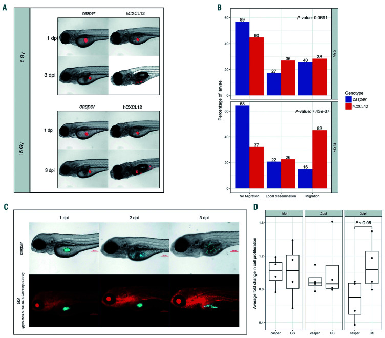
IMAGE

Figure 2.

ID
ZDB-IMAGE-201017-35
Source
Figures for Rajan et al., 2019
Image
Figure Caption

Figure 2.

Irradiation of human CXCL12-expressing transgenic zebrafish dramatically increases cell migration, while myeloid leukemia cells exhibit enhanced proliferation in the presence of CSF2/GM-CSF and KITLG/SCF. (A) Human Jurkat cells (which highly express CXCR4, the receptor for CXCL12) were xenografted into control casper and CXCL12-expressing larvae, which were further divided into two groups: one which received 15 Gy irradiation and the other no irradiation at 72 hours postfertilization (hpf). Larvae were screened for cell migration at 144 hpf. Representative images of control and CXCL12-expressing larvae that were not irradiated uooer panel). Representative images of control and CXCL12-expressing larvae following 15 Gy gamma irradiation (lower panel). (B) Quantification of cell migration was classified into “no migration,” “local dissemination” (dissemination within the yolk sac) and “migration” (distant migration beyond the yolk sac). Results represent three independent experiments. Numbers on the bar denote the total number of larvae per classification. (C) Representative images of zebrafish injected with CMK cells, a myeloid leukemia of Down syndrome (ML-DS) cell line. (D) Cell proliferation was quantified in transgenic larvae expressing GM-CSF/CSF2 and SCF/KITLG (GS fish) and casper controls following enzymatic digestion and dissociation at 1 day post-injection (dpi) (baseline), 2 dpi and 3 dpi. The analysis included fluorescence microscopy and cell counting. At 2 dpi there was a slight decrease in cell numbers in both transgenic and control larvae. By 3 dpi there was an increase in cell numbers in GS larvae, whereas cell numbers in control larvae decreased. Data presented represents four replicates with each dot in the bar graph denoting a single replicate. SCF/KITLG: stem cell factor/KIT ligand; GM-CSF/CSF2: granulocyte-monocyte colony-stimulating factor/colony-stimulating factor 2.

Acknowledgments
This image is the copyrighted work of the attributed author or publisher, and ZFIN has permission only to display this image to its users. Additional permissions should be obtained from the applicable author or publisher of the image. Full text @ Haematologica