
|
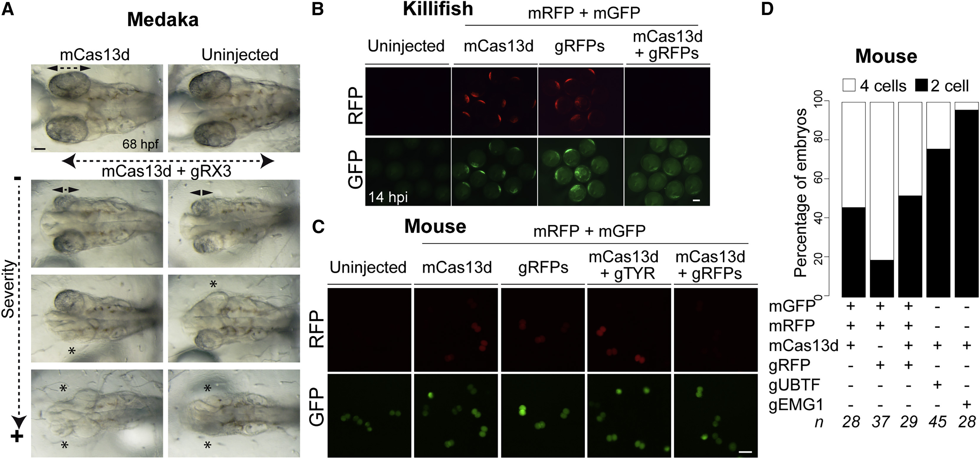
Fig. 5 CRISPR-RfxCas13d as RNA Targeting Tool in Different Animal Models (A) Representative pictures of medaka embryos at 68 hpf showing arrested eye development in embryos injected with mCas13d (450 pg) with gRNAs (1 ng) targeting rx3 mRNA (gRX3) compared with the embryos injected with mCas13d alone or uninjected embryos. Double arrows and asterisk indicate eye length and absence of eye, respectively (scale bar, 0.1 mm). (B) Fluorescence microscopy images of killifish embryos showing RFP and GFP intensity of uninjected embryos or after 14 hpi injected with rfp and gfp mRNAs, with or without mCas13d (250–350 pg/embryo) and/or gRNAs (300–400 pg/embryo) targeting rfp mRNA (scale bar, 0.1 mm). (C) Fluorescence microscopy images of mouse embryos 24 hpi (2 cells stage) with rfp and gfp mRNAs with or without mCas13d and/or gRNAs targeting rfp or unrelated (zebrafish tyr) mRNA (scale bar, 100 μm). (D) Stacked plots showing the percentage of mouse embryos at 2 or 4 cells stage 48 hpi with the indicated mRNA and/or gRNAs. The number of embryos (n) is indicated from at least two independent experiments.
Reprinted from Developmental Cell, 54(6), Kushawah, G., Hernandez-Huertas, L., Abugattas-Nuñez Del Prado, J., Martinez-Morales, J.R., DeVore, M.L., Hassan, H., Moreno-Sanchez, I., Tomas-Gallardo, L., Diaz-Moscoso, A., Monges, D.E., Guelfo, J.R., Theune, W.C., Brannan, E.O., Wang, W., Corbin, T.J., Moran, A.M., Sánchez Alvarado, A., Málaga-Trillo, E., Takacs, C.M., Bazzini, A.A., Moreno-Mateos, M.A., CRISPR-Cas13d Induces Efficient mRNA Knockdown in Animal Embryos, 805-817.e7, Copyright (2020) with permission from Elsevier. Full text @ Dev. Cell