
|
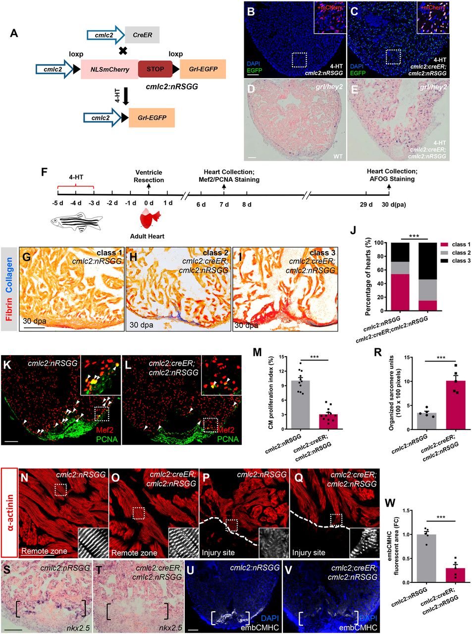
Fig. 3 Conditional grl induction in the adult myocardium impairs CM dedifferentiation and proliferation during regeneration. (A) Transgenic zebrafish used for inducible expression of Grl-EGFP in CMs. Tg(cmlc2:nRSGG) zebrafish were crossed with Tg(cmlc2:CreER) animals, which permitted Cre-mediated recombination to induce Grl in CMs after 5 µM 4-HT treatment. (B,C) Grl was induced in CMs of 4-HT-treated Tg(cmlc2:creER;cmlc2:nRSGG) hearts (C) but not Tg(cmlc2:nRSGG) hearts (B), as indicated by EGFP protein (green). Insets show higher-magnification images of the dashed boxes adding mCherry channel (red). (D,E) ISH analyses indicate the endogenous expression of grl in WT hearts (D) and the induced expression of grl in CMs of the whole ventricle in 4-HT-treated Tg(cmlc2:creER;cmlc2:nRSGG) animals (E) at 7 dpa. (F) Experimental design for PCNA and Mef2 immunostaining and fibrotic scar (AFOG) analysis after ventricular resection. 4-HT treatment was 5 µM 4-HT for bath treatment for 2 days. (G-I) Section images of 30 dpa ventricles of 4-HT-treated Tg(cmlc2:nRSGG) control fish (G) and 4-HT-treated Tg(cmlc2:creER;cmlc2:nRSGG) animals (H,I) stained with AFOG (blue for collagen, red for fibrin). Heart sections were scored as ‘class 1’, ‘class 2’ or ‘class 3’ according to the criteria described in Materials and Methods. (J) Quantification of regenerative status of ventricles in 4-HT-treated Tg(cmlc2:creER;cmlc2:nRSGG) (n=6; 129 sections) and Tg(cmlc2:nRSGG) control group (n=4; 84 sections) at 30 dpa. Histograms show the percentage of wounded hearts represented by each score for each group. ***P<0.001, Chi-square test. (K,L) Immunofluorescent section images of injured ventricles from 4-HT-treated Tg(cmlc2:nRSGG) control fish (K) and 4-HT-treated Tg(cmlc2:creER;cmlc2:nRSGG) animals (L) at 7 dpa, stained with anti-PCNA (green) and anti-Mef2 (red) antibodies. Insets show higher-magnification images of the dashed boxes. Arrowheads indicate proliferating CMs. (M) Quantification of CM proliferation indices in 7 dpa ventricles; n=11 (cmlc2:nRSGG), n=12 (cmlc2:creER;cmlc2:nRSGG); ***P<0.001, Student's t-test (unpaired, two-tailed). (N-Q) Confocal images of sections of injured ventricles from 4-HT-treated Tg(cmlc2:nRSGG) control fish (N,P) and 4-HT-treated Tg(cmlc2:creER;cmlc2:nRSGG) animals (O,Q) at 7 dpa, stained with anti-α-actinin antibody, showing regions of the remote zone (N and O) and injury site (P and Q). Insets show higher-magnification images of the dashed boxes. Dashed line indicates the apical edge of the regeneration. (R) Quantification of organized sarcomere units in α-actinin-labeled myocardial tissue (100×100 pixels) in injury border zone of 7 dpa ventricles from 4-HT-treated Tg(cmlc2:nRSGG) control fish and 4-HT-treated Tg(cmlc2:creER;cmlc2:nRSGG) animals (n=5); ***P<0.001, Student's t-test (unpaired, two-tailed). (S,T) ISH analyses of nkx2.5 expression in injured ventricles from 4-HT-treated Tg(cmlc2:nRSGG) control fish (S) and 4-HT-treated Tg(cmlc2:creER;cmlc2:nRSGG) animals (T) at 7 dpa (n=5). Brackets indicate injury site. (U,V) Fluorescent images of injured ventricles from 4-HT-treated Tg(cmlc2:nRSGG) control fish (U) and 4-HT-treated Tg(cmlc2:creER;cmlc2:nRSGG) animals (V) at 7 dpa, stained with embCMHC antibody (white). Brackets indicate injury site. (W) Quantification of embCMHC fluorescent area in injury border zone of 7 dpa ventricles from 4-HT-treated Tg(cmlc2:nRSGG) control fish and 4-HT-treated Tg(cmlc2:creER;cmlc2:nRSGG) animals. Data are relative to area in 4-HT-treated Tg(cmlc2:nRSGG) group (n=5); ***P<0.001, Student's t-test (unpaired, two-tailed). Data presents as mean±s.e.m. Scale bars: 100 µm (B-E,G-I,K,L,S-V); 50 µm (N-Q).