IMAGE

Figure 3

ID
ZDB-IMAGE-201003-98
Source
Figures for McCallum et al., 2020
Image
Figure Caption

Figure 3 Her4.3GFP transgenic line identifies cells with morphologies indicative of distinct subtypes of EGCs in the adult ENS.

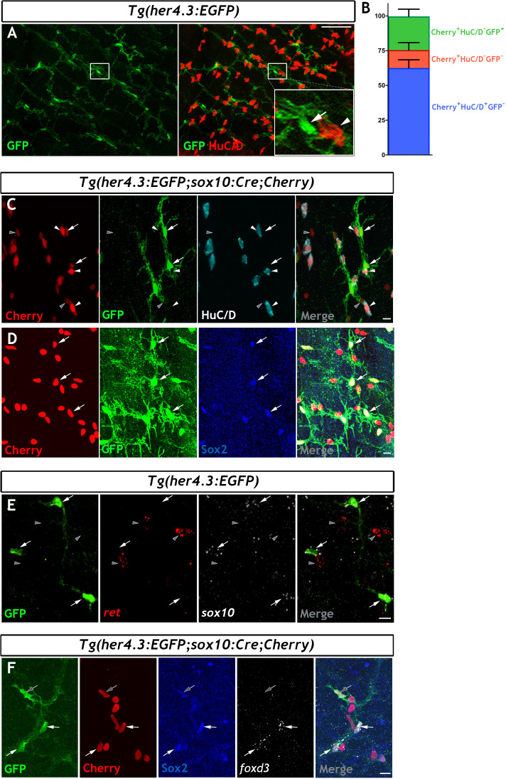
Immunohistochemistry of adult guts from of Tg(her4.3:EGFP) allow characterization the cellular morphology of GFP+ cells and comparisons to mammalian EGC subtypes (Boesmans et al., 2015). (A) GFP expressing cells (green) show close association with neurons, which express HuC/D in cell bodies and AcTu in cell processes (red) (n = 70). Inset shows high magnification view of boxed region, marking neurons (asterisks), GFP expression (arrowhead), and highly branched GFP expressing cellular processes (arrows). (B–E) Four distinct morphological cell types can be observed in Tg(her4.3:EGFP)+ cells: (B) GFP+ cells in the myenteric layer (arrowhead) with processes that appear to wrap around HuC/D+ cell bodies (red, asterisk), similar to Type I mammalian EGCs (inset), (C) GFP+ cells in the myenteric layer (arrowhead) with elongated processes (arrow) that follow AcTu+ neuronal processes (red), similar to Type II mammalian EGCs (inset), (D) GFP+ cells close to the mucosal layers (arrowhead), such as mucosal epithelia (ep, with DAPI highlighted nuclei in grey), similar to mammalian Type III EGCs (inset), and (E) Bipolar GFP+ cells within the muscle layers (arrowhead), associated with AcTu+ neuronal fibres (red, arrow), similar to Type IV mammalian EGCs (inset). Inset pictures adapted from Boesmans et al., 2015. All confocal images are max projections of short confocal stacks. Scale bars in merge panels: 50 µm (A) and 10 µm (B–E).

Acknowledgments
This image is the copyrighted work of the attributed author or publisher, and ZFIN has permission only to display this image to its users. Additional permissions should be obtained from the applicable author or publisher of the image. Full text @ Elife