
Image
|
Figure Caption
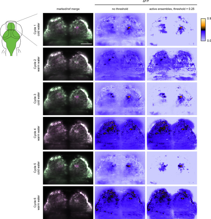
Figure 3—figure supplement 5.
Left image panels are merged marked (green) and reference erased (pseudo-colored magenta) images from the same Z position in the zebrafish pallium. Center image panels are the corresponding ΔF/F images. Right image panels are the corresponding images with threshold above 0.25 ΔF/F. Scale bar is 50 μm.
Acknowledgments
This image is the copyrighted work of the attributed author or publisher, and
ZFIN has permission only to display this image to its users.
Additional permissions should be obtained from the applicable author or publisher of the image.
Full text @ Elife