
Image
|
Figure Caption
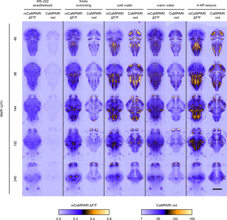
Figure 3—figure supplement 2.
Each image is an individual Z slice acquired at various depths from the brain of zebrafish larvae (4 to 5 dpf). Under each stimulus condition, images to the left are rsCaMPARI ΔF/F light sheet images from a fish expressing rsCaMPARI and images to the right are CaMPARI red confocal images from a different fish expressing CaMPARI. Scale bar is 200 μm.
Acknowledgments
This image is the copyrighted work of the attributed author or publisher, and
ZFIN has permission only to display this image to its users.
Additional permissions should be obtained from the applicable author or publisher of the image.
Full text @ Elife