
Image
|
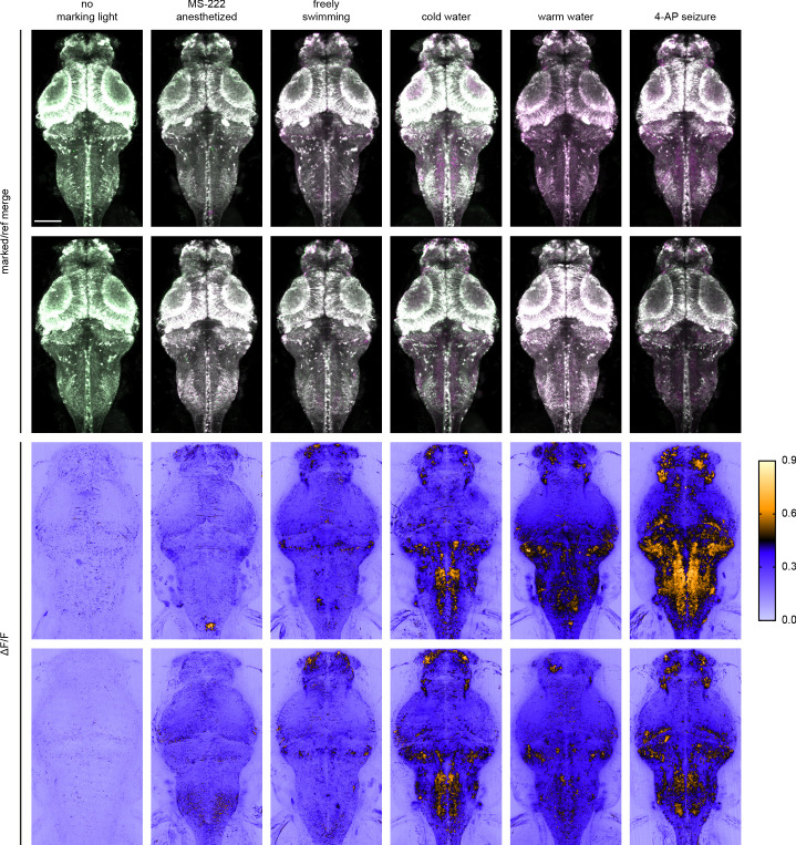
Figure Caption
Figure 3—figure supplement 1.
Each image is a maximum intensity Z projection of the entire brain from zebrafish larvae (4 to 5 dpf). Top half of images are merged marked and reference erased images, pseudo-colored green and magenta, respectively. Bottom half of images are corresponding ΔF/F images. Imaging conditions and brightness/contrast are identical to images shown in
Acknowledgments
This image is the copyrighted work of the attributed author or publisher, and
ZFIN has permission only to display this image to its users.
Additional permissions should be obtained from the applicable author or publisher of the image.
Full text @ Elife