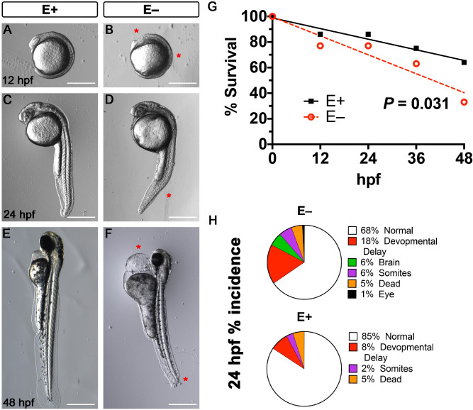
IMAGE

Figure 1

ID
ZDB-IMAGE-201003-143
Source
Figures for Head et al., 2020
Image
Figure Caption

Figure 1

Morphological abnormalities associated with VitE deficiency at 12-, 24- and 48 h post-fertilization. Representative bright field images of E+ and E− embryos showed normal development in E+ embryos and abnormalities in the E− embryos. At 12 hpf, E+ embryos (A) had defined somites and eyes, while E− embryos (B) showed abnormal somite formation and a reduced dorsal region where the eye is usually located. At 24 hpf, E+ embryos (C) had clearly defined eyes, heads and somites, while the E− embryos (D) had pericardial edema, less well-defined somites and notochord. At 48 hpf, E+ embryos (E) had extended fins and pigmentation throughout the body, while the E− embryos (F) experienced severe pericardial edema, stunted fin formation; some E− embryos experience errors in tail formation. (G) Early mortality, defined as nonviable beyond that time point, was increased in E− embryos at 12 hpf. By 48 hpf only about 35% of the original E− clutch survived with about 75% of E+ embryos surviving (P < 0.031) At 24 hpf (H), E− embryos that were alive experienced greater incidences of developmental delay, as well as morphological malformations that include brain, eye, and somites deformities. *Indicate delay or defects in E− embryos relative to E+ . Scale bar represents 500 m. Representative embryos are shown. Figure panels (AF) were generated with the BZ- × 700 microscope, processed with BZ-X Analyzer Software with image adjustments made with Adobe Photoshop v21.2.1.

Figure Data
Acknowledgments
This image is the copyrighted work of the attributed author or publisher, and ZFIN has permission only to display this image to its users. Additional permissions should be obtained from the applicable author or publisher of the image. Full text @ Sci. Rep.