Image
Figure Caption

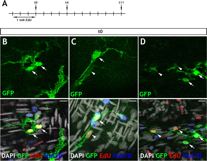
Figure 6—figure supplement 2. Adult <italic>Tg(her4.3:EGFP)</italic> cells take up EdU and appear in doublets.

(A) Schematic of experimental design: Immersion of 3 month old adult Tg(her4.3:EGFP) zebrafish in 1 mM EdU pulse for three days was followed by a return to normal zebrafish water. Animals were then culled after chase periods of 0 days (t0), 4 days (t4) or 11 days (t11) and analysed for EdU incorporation (t0 n = 6, t4 n = 5, t11 n = 5). (B–D) At 0 days chase, the majority of EdU labelled GFP+ (yellow) cells are found in doublets (two labelled cells in close proximity). These cells are either: (B) both expressing high levels of GFP (green, arrows), (C) appear with one high GFP expressing cell (arrow) and one low GFP expressing cell (arrowhead), (D) in larger groupings, where EdU labelling is associated with cells exhibiting lower levels of GFP expression (arrowhead) and not observed in high GFP expressing cells (arrows). Confocal images are max projections of short confocal stacks. Scale bars in merge panels: 10 µm (B–D).

Acknowledgments
This image is the copyrighted work of the attributed author or publisher, and ZFIN has permission only to display this image to its users. Additional permissions should be obtained from the applicable author or publisher of the image. Full text @ Elife