
|
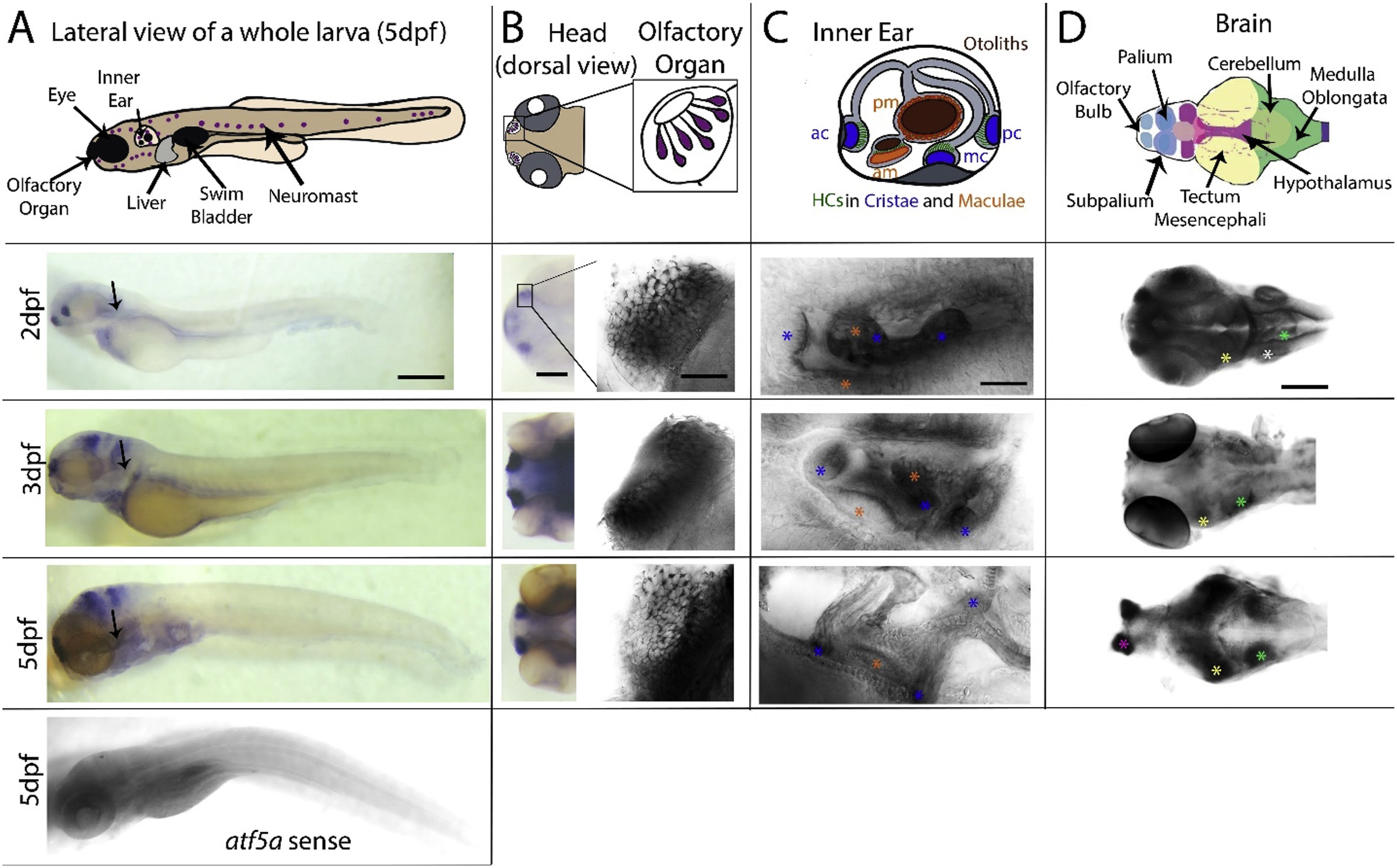
Fig. 3 Larval expression of atf5a. (A) Schematic of a whole larva at 5dpf (top panel) locating the major sensory organs (eyes, olfactory organs and neuromasts (NMs) of the superficial sensory lateral line organ, LL) as well as the swim bladder and the liver. Whole larvae were hybridized with an antisense RNA probe at 2, 3 and 5dpf (3 top panels) and with a sense probe at 5dpf (lower panel). Black arrows indicate the developing liver. (B) Dorsal views of the rostral part of the larva (left column) showing strong atf5a expression in the olfactory epithelia (OE) of olfactory organs. OE are shown at higher magnification (right column). (C) Lateral views of the inner ear (schematized in the top panel highlighting all sensory epithelia with hair cells (HCs, green): anterior (am) and posterior maculae (pm, orange) sitting underneath otoliths (brown), and anterior (ac), median (mc) and posterior cristae (pc, in blue) sitting at the end of each corresponding semicircular canal. The atf5a gene is expressed in maculae (orange asterisks) and cristae (blue asterisks) at all stages. (D) Dorsal views of the head showing the different brain regions. Schematic adapted from Torres-Hernandez et al. (2016) (top panel) depicting telencephalon (blue), diencephalon (magenta), mesencephalon (yellow) and metencephalon (green). At 2dpf and 3dpf (2nd and third panels) whole larva had visible atf5a expression in the inner ear (white asterisk), the tecta mesencephali (yellow asterisks), and the metencephalic medulla oblongata (green asterisks). At 5dpf, dissected brains showed strong expression in the sub-mentioned regions and in the olfactory bulbs (OBs, pink asterisks). Scale bars: in A = 200 μm, in B (left column) = 75 μm, in B (right column) = 25 μm, in C = 50 μm, and in D = 150 μm.
Reprinted from Gene expression patterns : GEP, 37, Rodríguez-Morales, R., Vélez-Negrón, V., Torrado-Tapias, A., Varshney, G., Behra, M., Expression patterns of activating transcription factor 5 (atf5a and atf5b) in zebrafish, 119126, Copyright (2020) with permission from Elsevier. Full text @ Gene Expr. Patterns