
|
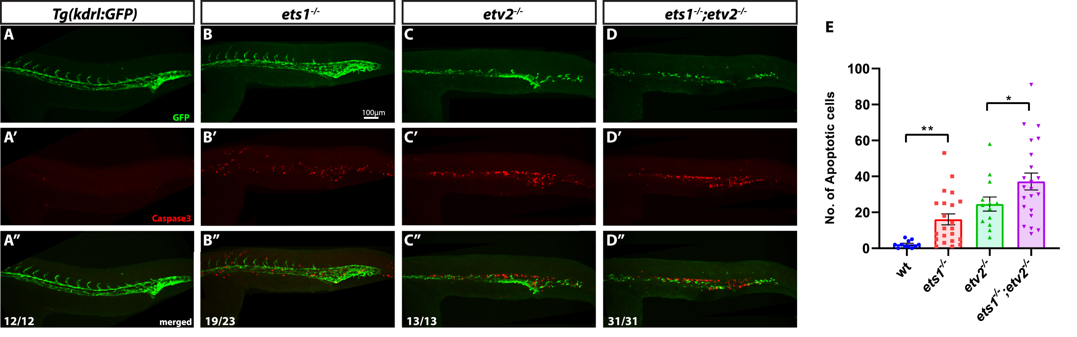
Fig. 4 Ets1 promotes endothelial cell survival in axial vessels. (A-D″) Tg(kdrl:GFP), ets1−/−, etv2−/− and ets1−/−; etv2−/−embryos at 24 hpf immuno-stained for apoptosis marker Cleaved Caspase3 and vascular GFP. Note the normal vasculature in ets1−/−embryos (B) despite extensive Caspase 3 staining in the axial vessels (B′). Both etv2−/− (C-C″) and ets1−/−; etv2−/−embryos (D-D″) exhibit severe vascular defects and extensive apoptosis in the trunk region. (E) Quantification of apoptotic cells within trunk vasculature in 24 hpf embryos. Note the increase in apoptotic cells in the ets1−/−; etv2−/− embryos compared to etv2−/− embryos. ∗P < 0.05, ∗∗P < 0.01; horizontal bars represent mean, error bars represent ± SEM.
Reprinted from Developmental Biology, 465(1), Chetty, S.C., Sumanas, S., Ets1 functions partially redundantly with Etv2 to promote embryonic vasculogenesis and angiogenesis in zebrafish, 11-22, Copyright (2020) with permission from Elsevier. Full text @ Dev. Biol.