Image
Figure Caption

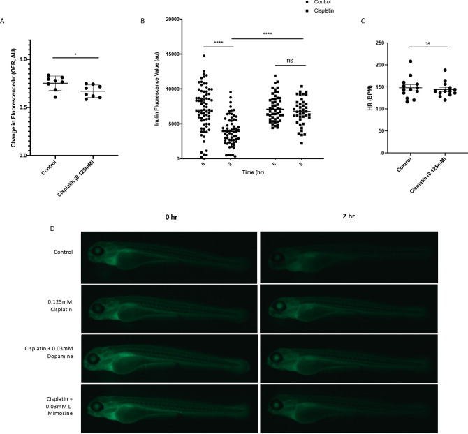
Figure 5—figure supplement 1. Optimization of the experimental detection of cisplatin-induced decreases in glomerular filtration rate (GFR) in zebrafish larvae.

Casper zebrafish larvae were treated at 72 hr post-fertilization (hpf) with either vehicle control or 0.125 mM cisplatin for 24 hr. Larvae were rinsed, then injected via the common cardinal vein with FITC-inulin. (A) Inulin-injected larvae were imaged immediately after injection and 2 hr after injection. Larval fluorescence was measured using ImageJ software using a region of interest (ROI) superimposed on the tail of each larvae. The change in fluorescence between the two time points (a relative measure of GFR) was calculated, and is displayed in arbitrary units (AU). Each data point represents the change in fluorescence of an individual larvae. Error bars represent SEM. *=p<0.05, as per two-tailed student’s t-test. N=2, at least four larvae/treatment group/experiment. (B) Larvae were treated as in A), but were measured for overall larval fluorescence immediately after injection and 2 hr after injection using a Biosorter. Results display raw larval fluorescence values. Each data point represents an individual larvae at each time point. ****=p<0.001, as per two-way ANOVA with a tukey post-test. N=1, with at least 50 larvae/treatment group. (C) Heart rate (HR) was measured in larvae treated as in A), to determine if changes in HR had an effect on the measured GFR. There was no significant difference between HR regardless of treatment, as per two-tailed student’s t-test. Each data point represents an individual larvae. N=2, with at least six larvae/treatment group. (D) Representative larvae from the experiment described in the main text, and found in Figure 5a. Briefly, casper zebrafish larvae were treated at 60 hpf with either vehicle control or the indicated protective agents. At 72 hpf, larvae were rinsed then treated with either vehicle control or 0.125 mM cisplatin for 24 hr. Larvae were then injected via the common cardinal vein with FITC-inulin, then were imaged using a Zeiss SteREO Discovery.V20 dissecting microscope.

Acknowledgments
This image is the copyrighted work of the attributed author or publisher, and ZFIN has permission only to display this image to its users. Additional permissions should be obtained from the applicable author or publisher of the image. Full text @ Elife