IMAGE

Figure 4.

ID
ZDB-IMAGE-200904-22
Source
Figures for Hu et al., 2020
Image
Figure Caption

Figure 4.

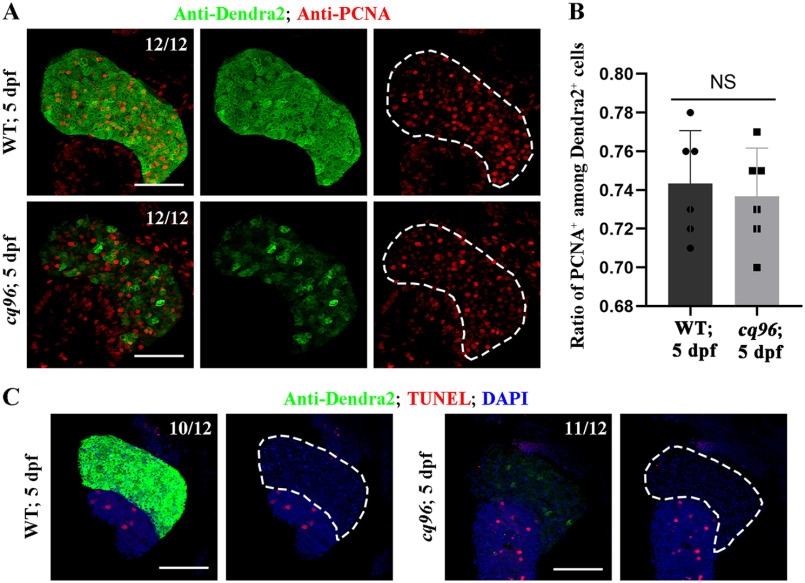
Loss of Rbm15 has no effect on hepatic proliferation and apoptosis.A, antibody staining results of PCNA and Dendra2 showing hepatocytes outside of the G0 phase at 5 dpf. B, quantification of the percentage of the PCNA+ among Dendra2+ cells at 5 dpf in WT and rbm15−/−. TUNEL assay images showing the apoptosis of liver in WT and rbm15−/− at 5 dpf. Numbers indicate the proportion of larvae exhibiting the expression shown. NS, not significantly different; scale bars, 100 μm; error bars, S.D.

Acknowledgments
This image is the copyrighted work of the attributed author or publisher, and ZFIN has permission only to display this image to its users. Additional permissions should be obtained from the applicable author or publisher of the image. Full text @ J. Biol. Chem.