IMAGE

Figure 4

ID
ZDB-IMAGE-200829-51
Source
Figures for Bernut et al., 2020
Image
Figure Caption

Figure 4

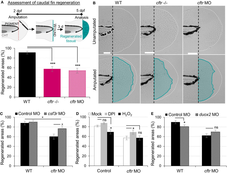
Neutrophilic response in CF hampers tissue repair in vivo. (A,B) Tail fin regeneration assessment in WT, cftr –/– and cftr MO zebrafish. Two dpf embryos were tail fin amputated then the potential of tail fin regeneration is evaluated by measuring regenerated fin areas at 3 dpi (blue). (A) Measurement of regenerated fin areas (n = 30, one-way ANOVA with Dunnett's post-test). (B) Representative imaging of injured tail fin at 3 dpi (scale bars, 200 μm). (C) Selective ablation of neutrophils has been carried out in WT and CF animals by injecting csf3r-MO. Measurement of regenerated fin areas in the presence or absence of neutrophils (n = 21, one-way ANOVA with Tukey post-test). (D)TgBAC(mpx:EGFP)i114 larvae were pretreated with DPI or H2O2 prior to tail fin amputation procedures, then injured and immediately put back in treatments for 4 h. Regenerated fin areas are measured at 3 dpi (n = 21, one-way ANOVA with Dunnett's post-test). (E) Inhibition of NADPH oxidase activity by injecting duox2-MO has been carried out in WT and CF animals. Embryos were tail fin injured and regenerated fin areas measured at 3 dpi (n = 21, two-tailed Bonferroni t-test).

Figure Data
Acknowledgments
This image is the copyrighted work of the attributed author or publisher, and ZFIN has permission only to display this image to its users. Additional permissions should be obtained from the applicable author or publisher of the image. Full text @ Front Immunol