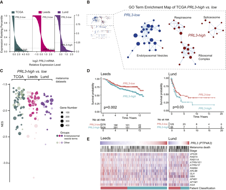
IMAGE

Figure 7

ID
ZDB-IMAGE-200824-7
Source
Figures for Johansson et al., 2020
Image
Figure Caption

Figure 7

A PRL3-High, Endolysosomal Gene-Low Signature in Human Melanoma Samples

(A) Patient samples ranked by PRL3 RNA expression. PRL3-high and PRL3-low subgroups were defined with a 10% threshold for TCGA (n = 366) and Leeds (n = 703) and 25% threshold for the Lund dataset (stage III patients, n = 124).

(B) GO enrichment analysis (cellular compartment) of PRL3 subgroups in TCGA (Cytoscape). Node radius represents GO term gene counts. Spokes represent shared gene counts between terms.

(C) Bubble plot of GO cellular components in PRL3-high versusPRL3-low melanomas. Over-representation analysis (FDR < 0.05). NES, normalized enrichment score.

(D) Kaplan Meier survival curves of PRL3-high versus PRL3-low melanomas in the Leeds and Lund cohorts Leeds dataset: p = 0.002; Lund dataset: p = 0.03; logrank tests.

(E) Heatmap of endolysosomal genes ranked by PRL3 expression (mean-centered): high (red), low (blue). Melanoma deaths are indicated in black; stages are indicated by shades of gray, darker shades represent higher stages. Patient classification: PRL3-low (red), PRL3-high (green), as in (D).

See also Figure S6; Table S6.

Acknowledgments
This image is the copyrighted work of the attributed author or publisher, and ZFIN has permission only to display this image to its users. Additional permissions should be obtained from the applicable author or publisher of the image.

Reprinted from Developmental Cell, 54(3), Johansson, J.A., Marie, K.L., Lu, Y., Brombin, A., Santoriello, C., Zeng, Z., Zich, J., Gautier, P., von Kriegsheim, A., Brunsdon, H., Wheeler, A.P., Dreger, M., Houston, D.R., Dooley, C.M., Sims, A.H., Busch-Nentwich, E.M., Zon, L.I., Illingworth, R.S., Patton, E.E., PRL3-DDX21 Transcriptional Control of Endolysosomal Genes Restricts Melanocyte Stem Cell Differentiation, 317-332.e9, Copyright (2020) with permission from Elsevier. Full text @ Dev. Cell