
|
Figure 1
Prl3a Is an Inhibitor of Melanocyte Regeneration in Zebrafish
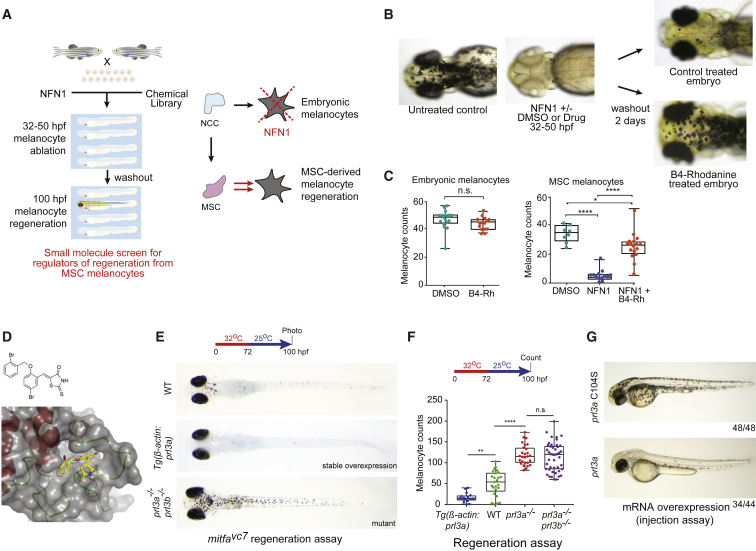
(A) Schematic of a small-molecule screen for regulators of MSC-derived melanocytes in zebrafish. MSC, melanocyte stem cell; NCC, neural crest cells.
(B) Images of zebrafish embryos treated with NFN1 ± DMSO or B4-Rh and after washout.
(C) Quantification of zebrafish melanocytes during normal development (n.s., not significant, Student’s t test) or in a NFN1-regeneration assay (ANOVA using Tukey’s analysis; ∗p value = 0.0131; ∗∗∗∗p ≤ 0.0001).
(D) Predicted binding of B4-Rh (yellow sticks) in the NMR model of PRL3 (gray transparent surface and secondary structure; red, helix; green, loop). Purple-dashed line: predicted hydrogen bond to E50. All other protein-ligand interactions are apolar. The ligand sits in a hydrophobic pocket formed by residues: V48, C49, W68, P69, A74, P75, P77, V80, A111, V113, and the methylene groups of the side chain of Q145. These residues, and E50, are conserved in zebrafish Prl3a.
(E and F) (E) Images and (F) quantification of wild type,
(G) RNA overexpression of
See also
Reprinted from Developmental Cell, 54(3), Johansson, J.A., Marie, K.L., Lu, Y., Brombin, A., Santoriello, C., Zeng, Z., Zich, J., Gautier, P., von Kriegsheim, A., Brunsdon, H., Wheeler, A.P., Dreger, M., Houston, D.R., Dooley, C.M., Sims, A.H., Busch-Nentwich, E.M., Zon, L.I., Illingworth, R.S., Patton, E.E., PRL3-DDX21 Transcriptional Control of Endolysosomal Genes Restricts Melanocyte Stem Cell Differentiation, 317-332.e9, Copyright (2020) with permission from Elsevier. Full text @ Dev. Cell