
Image
|
Figure Caption
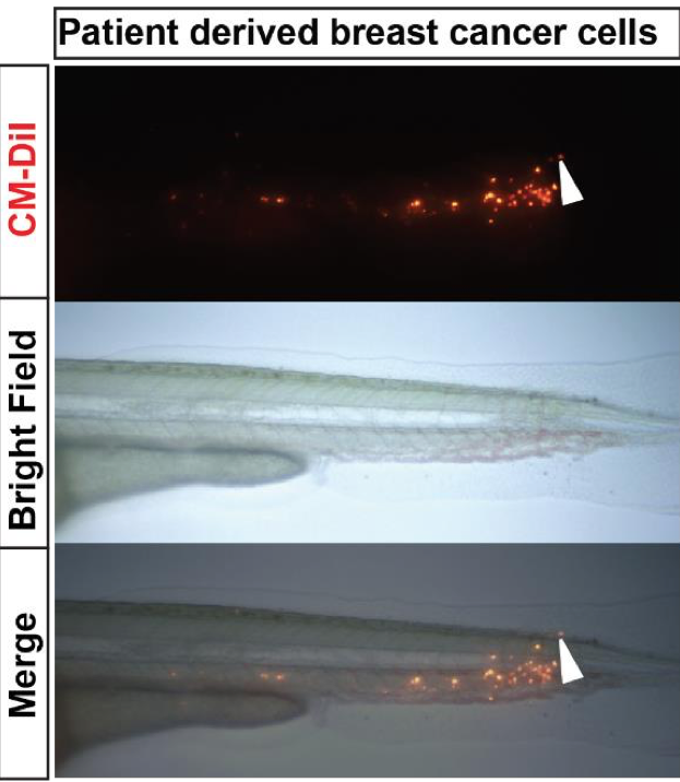
Fig. S5
Tail invasion of primary breast tumor cells. Primary tumor cells (top) were labeled with CM-DiI and injected in the eZXM. Imaging of the tail part of at 1 dpi (bright field image, middle), revealed invasion of the tail fin fold region by a single cell (white arrowhead).
Acknowledgments
This image is the copyrighted work of the attributed author or publisher, and
ZFIN has permission only to display this image to its users.
Additional permissions should be obtained from the applicable author or publisher of the image.
Full text @ Sci. Rep.