
|
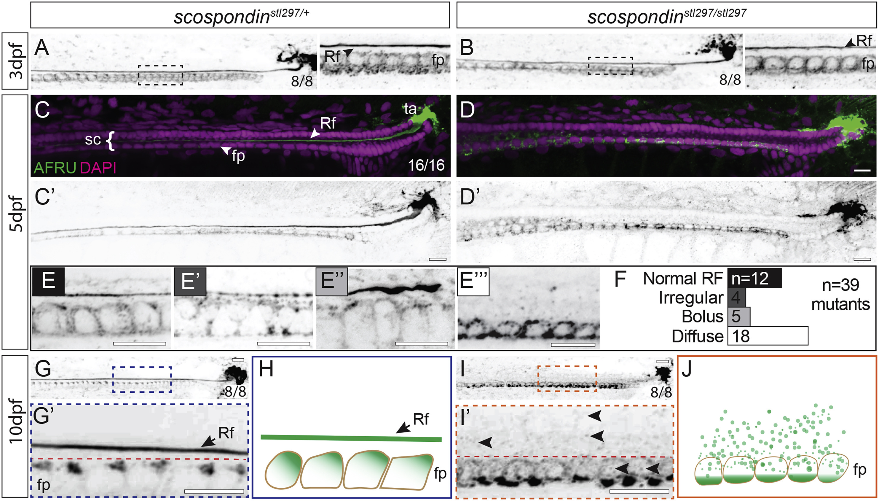
Fig. 2 Disassembly of the Reissner Fiber due to Defects in Secretion from the Floor Plate Is Correlated with the Onset of Axial Curvatures in Zebrafish Maximal Z projections of confocal stacks of the caudal region of the tail and spinal cord immunostained against the Reissner fiber in scospondinstl297/+ and scospondinstl297/stl297 mutants at 3 dpf (A and B), 5 dpf (C–F), and 10 dpf (G, G’, I, and I’). Rf, Reissner fiber; fp, floor plate; sc, spinal cord; ta, terminal ampulla. (A and B) At 3 dpf, both heterozygotes (A) and mutants (B) have an assembled Reissner fiber (8/8 each genotype). Insets to the right of (A) and (B) highlight a magnified region (dashed box). (C–E''') Pseudocolored merge from maximal Z projections of confocal stacks showing Reissner fiber (green) and DAPI (magenta) marking nuclei (C and D) and inverted grayscale image of Reissner fiber at 5 dpf (C’ and D’). We observed the Reissner fiber in all heterozygous mutants (100%; n = 16; C), but the scospondinstl297/stl297 showed some with the fiber (31%; n = 39) and some with a fiber in various stages of disassembly (69%; n = 39; D). We classified Reissner fiber staining as normal (E), irregular (E’), bolus (E’’), or diffuse (E’’’) and counted the number of mutants in each class (F). Scale bars, 10 μm. (G and I) At 10 dpf, we observed the Reissner fiber in all heterozygous controls (G), which was completely lost in the mutants (I; 100%; n = 7 and 8, respectively). Scale bars, 10 μm. (H and J) Schematic representation of Reissner material localization at 10 dpf. In wild-type or heterozygous animals, Reissner material localizes to the apical surface of floor plate cells and is assembled into a Reissner fiber (H). In scospondinstl297/stl297 mutants, the Reissner fiber is missing and Reissner material localizes at the basal portion of the floor plate (J). See also Figures S1 and S2.