
|
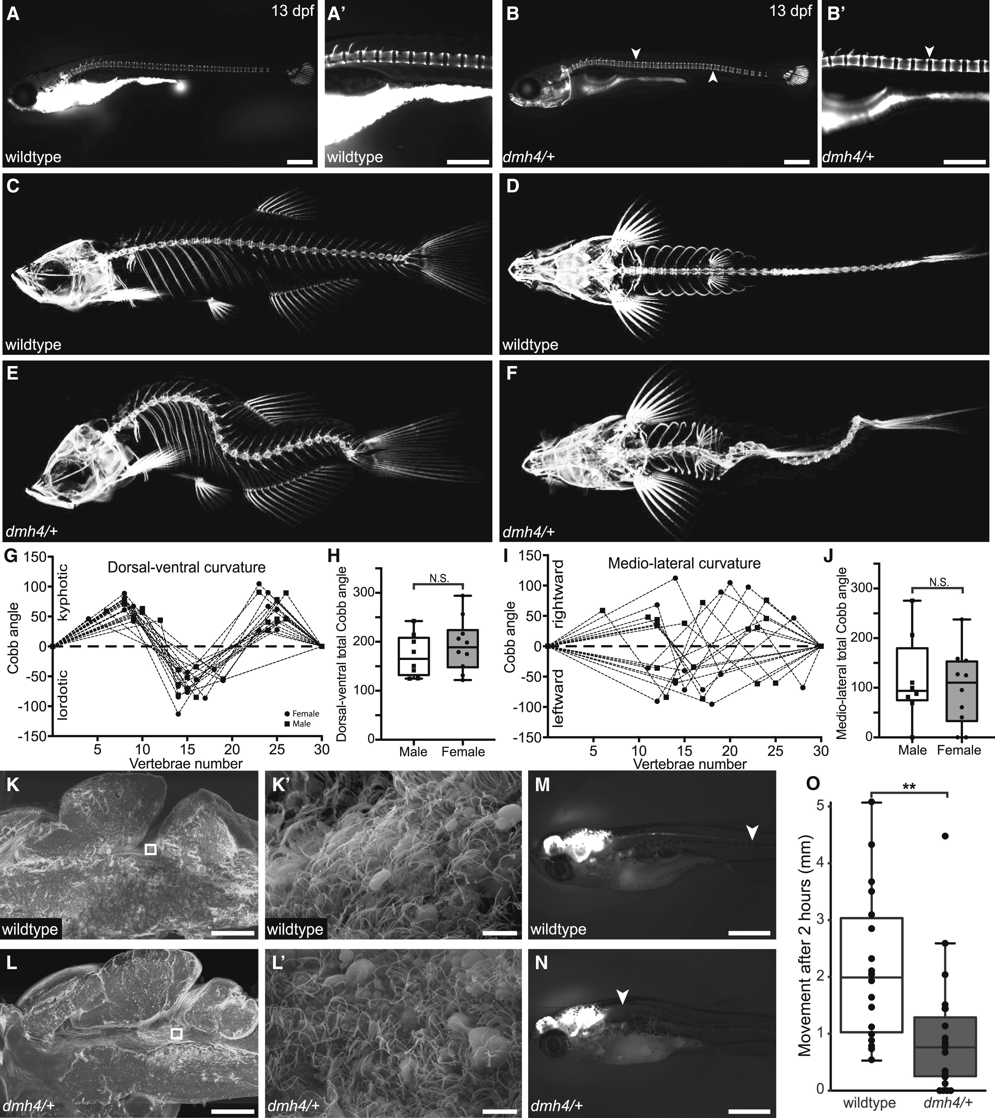
Fig. 1 Idiopathic-like Scoliosis in dmh4/+ Zebrafish Is Associated with Compromised CSF Flow in the Absence of Obvious Motile Cilia Defects (A and B) Calcein stains of wild-type (A and A’; N = 3; n = 24) and dmh4/+ (B and B’; N = 3; n = 29) larva at 13 dpf (6.5 mm standard length). Note the onset of spinal curvatures in dmh4/+ zebrafish (arrowheads) in the absence of congenital vertebral malformations. Scale bars represent 500 μm. (C–F) Lateral (C and E) and dorsal (D and F) projections of three-dimensional microCTs for adult wild-type (C and D) and dmh4/+ (E and F) fish. (G–J) Quantification of curve severity, direction, and position along dorsal-ventral (G) and mediolateral (I) axes. Dashed lines indicate a Cobb angle of 0°, as per a wild-type spine. Data points for male and female fish are indicated as squares and circles, respectively. Combined total Cobb angle in dorsal-ventral (H; p = 0.4) and medio-lateral (J; p = 0.67) axes for males and females is shown. (K and L) Representative SEM images of wild-type (K; N = 3; n = 7) and dmh4/+ (L; N = 3; n = 10) bisected brains at 3 months of age. Scale bars represent 200 μm. Squares indicate location of higher magnification images (K’ and L’). Scale bars represent 5 μm. (M–O) Representative images (M and N) and quantification (O) of bulk CSF movement along the spinal cord of 20-dpf wild-type (M; n = 18) and dmh4/+ (N; n = 18) larva, 2 h after injection of reporter dye into brain ventricles. p = 0.0038. Arrowheads indicate movement of dye. Scale bars represent 1 mm. See also Figure S1.