
|
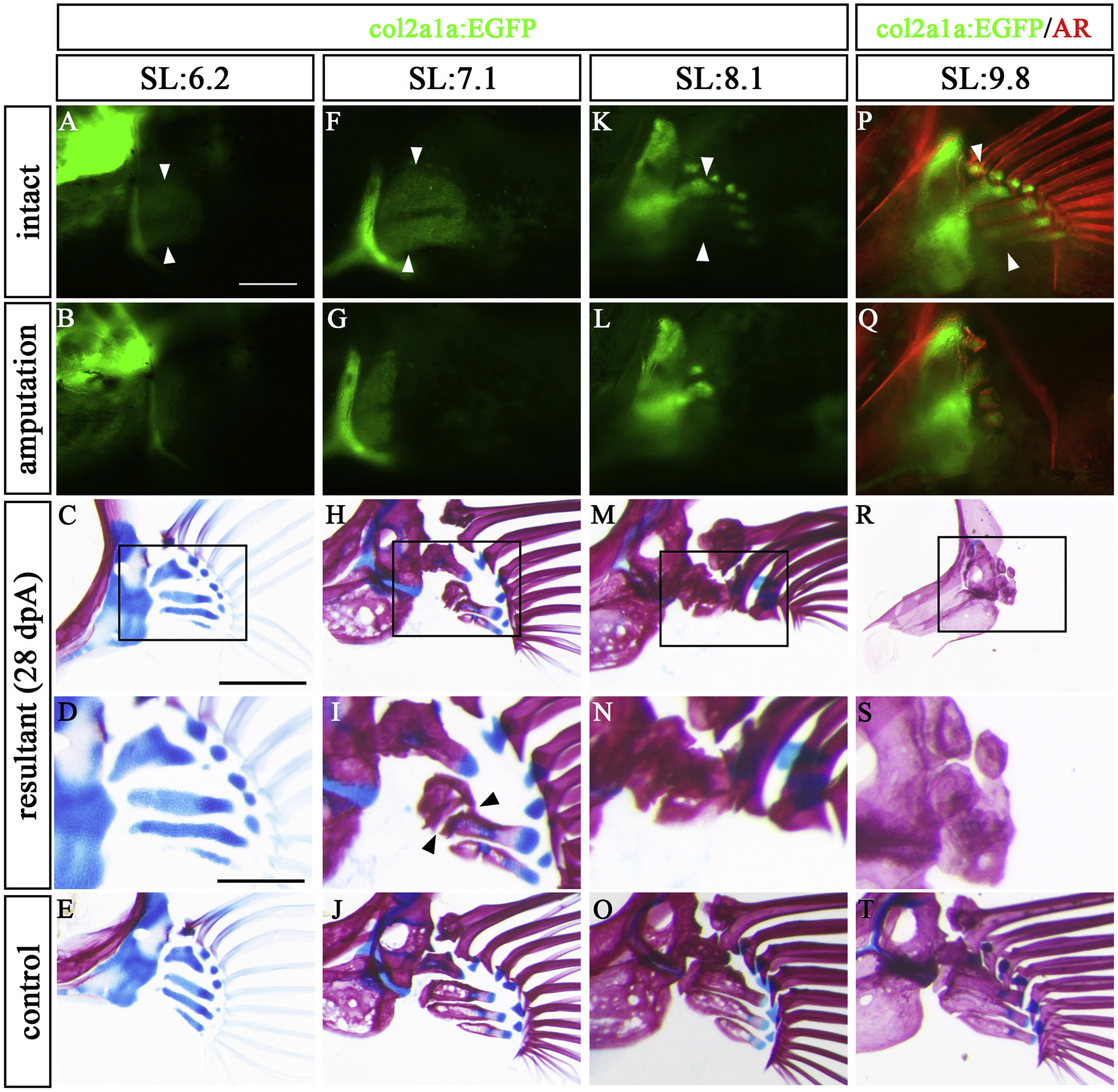
Fig. 2 Regeneration state differs by developmental stage. Developing pectoral fins in col2a1a:EGFP were amputated, and the fish were kept for 28 days. Pectoral fins of col2a1a:EGFP fish with 6.2 mm SL (A–E), 7.1 mm SL (F–J), and 8.1 mm SL (K–O) were amputated. (P–T) col2a1a:EGFP pectoral fin of 9.8 mm SL zebrafish was stained with AR and amputated. (A, F, K, P) Intact pectoral fins before amputation. (B, G, L, Q) Post-amputation pectoral fins at 0 dpA. (C, H, M, R) At 28 dpA, pectoral fin skeletons stained with AR and AB. (D, I, N, S) Magnified views of C, H, M, and R, respectively. (E, J, O, T) Control pectoral fins on the contralateral side of the 28 dpA specimens. These pictures are flipped horizontally. White arrowheads indicate amputation site. Black arrowheads indicate the site of disconnection between distal and proximal prs. Scale bars: 200 μm in A, C, 100 μm in D.
Reprinted from Developmental Biology, 463(2), Yoshida, K., Kawakami, K., Abe, G., Tamura, K., Zebrafish can regenerate endoskeleton in larval pectoral fin but the regenerative ability declines, 110-123, Copyright (2020) with permission from Elsevier. Full text @ Dev. Biol.