
|
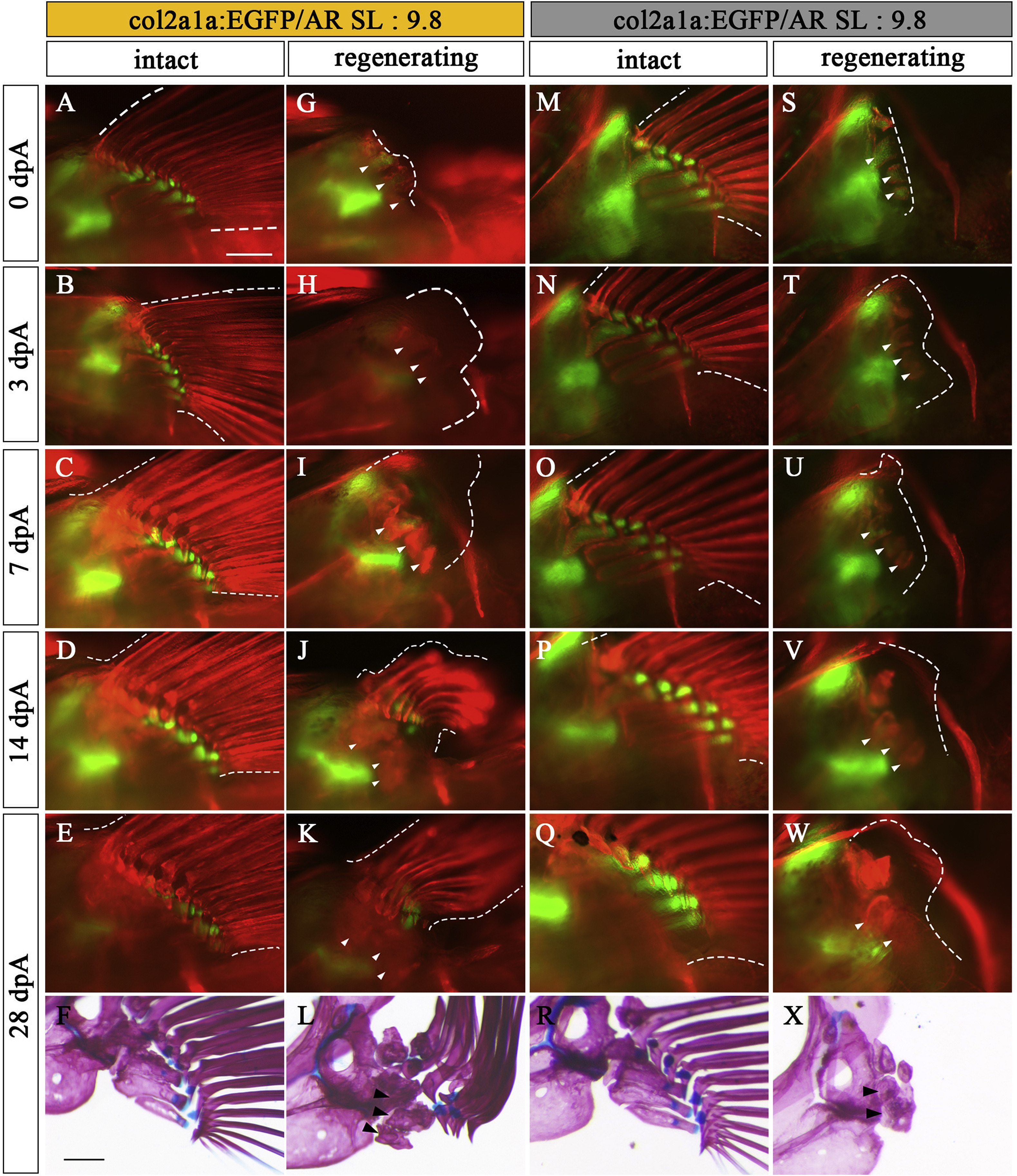
Fig. 5 Regeneration process of Type 2 and Type 3. Pectoral fins of col2a1a:EGFP specimens (SL: 9.8) showing typical Type 2 (A–L) and Type 3 (M–X) phenotypes. (A, M) Intact pectoral fins before amputation. (B-E, N-Q) Contralateral unamputated pectoral fins at 3 dpA (B, N), 7 dpA (C, O), 14 dpA (D, P), and 28 dpA (E, Q). These pictures are flipped horizontally. (G-K, S–W) Amputated pectoral fins at 0 dpA (G, S), 3 dpA (H, T), 7 dpA (I, U), 14 dpA (J, V), and 28 dpA (K, W). White arrowheads in U, V, and W indicate newly formed pr. (F, L, R, X) Pectoral fin skeleton of each sample stained with AR and AB at 28 dpA; L and X show typical Type 2 (L) and Type 3 (X) phenotypes, respectively. White dotted lines show the circumference of the pectoral fin. White arrowheads and black arrowheads show remnants of prs in pectoral fins. Scale bars: 200 μm.
Reprinted from Developmental Biology, 463(2), Yoshida, K., Kawakami, K., Abe, G., Tamura, K., Zebrafish can regenerate endoskeleton in larval pectoral fin but the regenerative ability declines, 110-123, Copyright (2020) with permission from Elsevier. Full text @ Dev. Biol.