
|
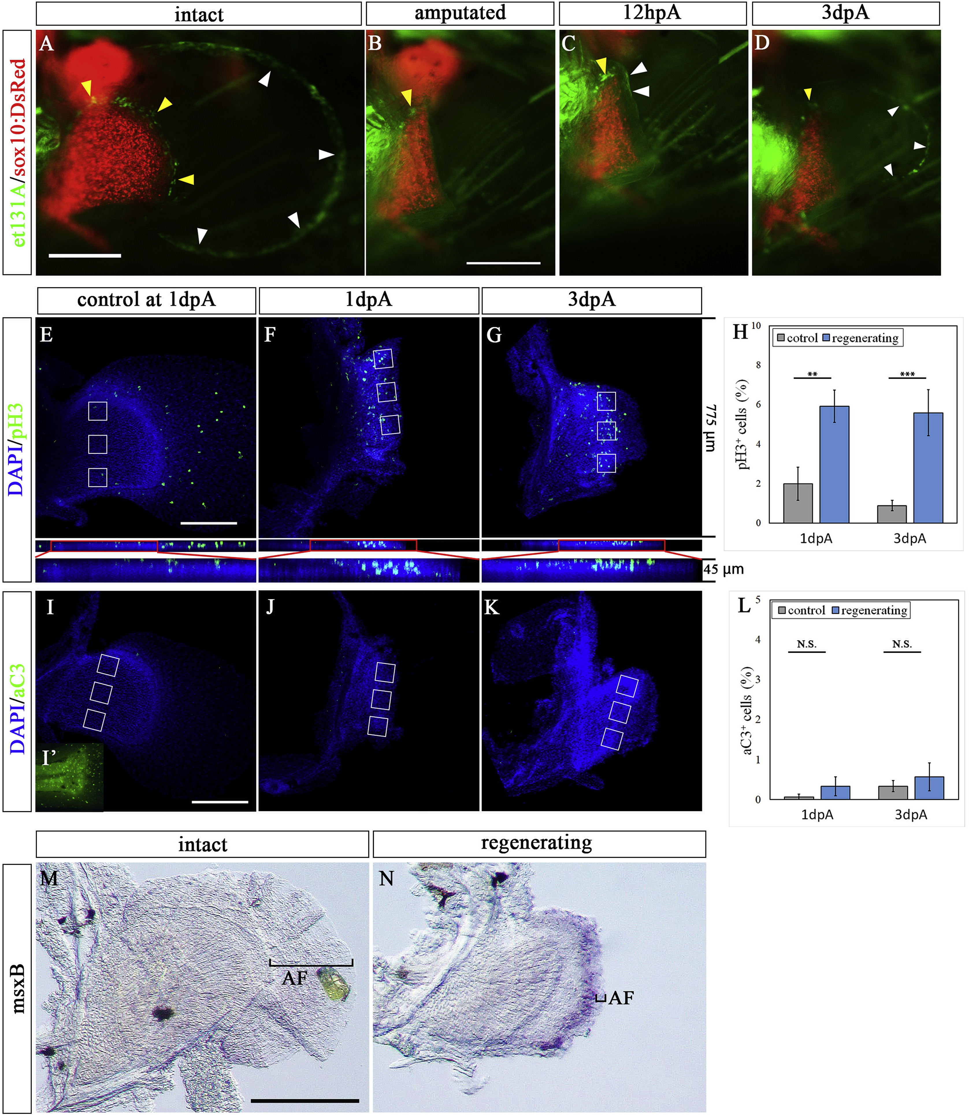
Fig. 6 The expression pattern of et131A, phospho-histone-H3, active Caspase 3, and msxB in amputated pectoral fin. (A–D) Pectoral fin of et131A;sox10:DsRed zebrafish. (A) Pre-amputation pectoral fin. (B–D) Amputated fin at 0 dpA (B), 12 hpA (C), and 3 dpA (D). White and yellow arrowheads indicate expression of UAS:EGFP of et131A in the epithelium and mesenchyme in the pectoral fin, respectively. (E–G) Expression pattern of phospho-histone-H3 in intact and regenerating pectoral fin; control contralateral fin at 1 dpA (E), amputated fin at 1 dpA (F) and at 3 dpA (G). Lower panels in E-G show compacted Z-stack images. (H) The proportion of pH3-positive cells near the amputation plane. This proportion was calculated from the number of pH3-positive cells in 3 regions of 100 cells in the areas enclosed in white boxes. White boxes in the control fins were positioned on the regions corresponding to those in the amputated fins where the pH3 signals were measured. Error bars are the mean SEM. (I–K) Expression pattern of active Caspase3 (aC3) in intact and regenerating pectoral fin with control contralateral fin at 1 dpA (I) and tail region of the same sample as the positive control of staining (I′), and amputated fin at 1 dpA (J) and at 3 dpA (K). (L) The proportion of aC3-positive cells near the amputation plane. This proportion was calculated from the number of aC3-positive cells in three regions of 100 cells in the areas enclosed in white boxes. White boxes in the control fins were positioned on the regions corresponding to those where the aC3 signals were measured in the amputated fins. Error bars indicate mean SEM. (MaC3-N) Expression pattern of msxB in intact contralateral (M) and regenerating (N) pectoral fins at 3 dpA. Black brackets indicate the area of apical fold (AF). The photo in M is horizontally flipped. Mean values ± SEM, ∗P < 0.05, ∗∗∗P < 0.001. Scale bars: 200 μm in A, E, and I and 100 μm in M.
Reprinted from Developmental Biology, 463(2), Yoshida, K., Kawakami, K., Abe, G., Tamura, K., Zebrafish can regenerate endoskeleton in larval pectoral fin but the regenerative ability declines, 110-123, Copyright (2020) with permission from Elsevier. Full text @ Dev. Biol.