
Image
|
Figure Caption
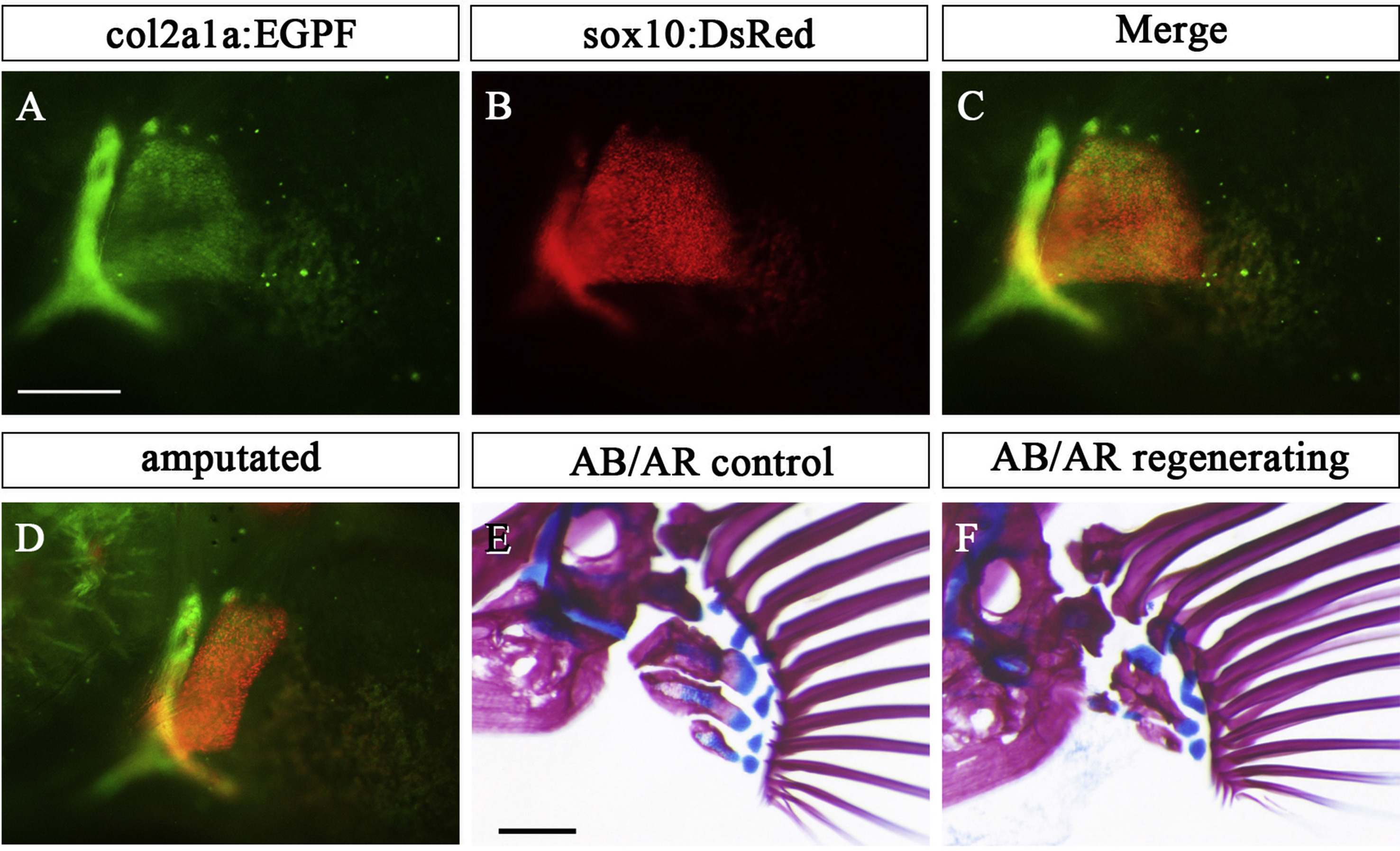
Fig. s2 Endoskeleton of pectoral fin in col2a1a:EGFP;sox10:DsRed double transgenic fish. (A-C) Expression pattern of col2a1a:EGFP (A), sox10:DsRed (B), and merged view (C) in the chondrocytes of the pectoral fin. (D) Amputated pectoral fin of col2a1a:EGFP; sox10:DsRed double transgenic fish. (E, F) Pectoral fin skeletons in col2a1a:EGFP; sox10:DsRed double transgenic fish stained with AR and AB at 28 dpA: contralateral control (E) and regenerated (F) pectoral fins. Scale bar: 200 μm in A, D and E.
Acknowledgments
This image is the copyrighted work of the attributed author or publisher, and
ZFIN has permission only to display this image to its users.
Additional permissions should be obtained from the applicable author or publisher of the image.
Reprinted from Developmental Biology, 463(2), Yoshida, K., Kawakami, K., Abe, G., Tamura, K., Zebrafish can regenerate endoskeleton in larval pectoral fin but the regenerative ability declines, 110-123, Copyright (2020) with permission from Elsevier. Full text @ Dev. Biol.